Download
1 / 37
671 likes | 1.97k Vues
Bacterial Skin Infections. Professor Sudheer Kher. Learning Objectives. Enumerate the microbes causing skin infections. Describe the characteristic clinical manifestations methods of laboratory diagnosis principles of management methods of prevention of each of the infections listed.

E N D
Bacterial Skin Infections Professor Sudheer Kher
Learning Objectives • Enumerate the microbes causing skin infections. • Describe the • characteristic clinical manifestations • methods of laboratory diagnosis • principles of management • methods of prevention of each of the infections listed.
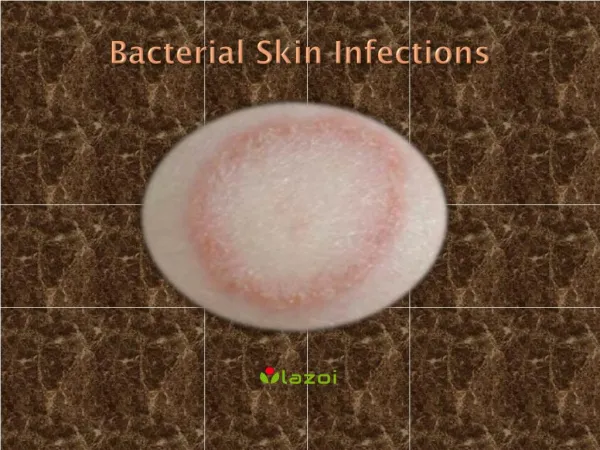
The Skin Definition Skin is largest organ of body. Maintains homeostasis, protects underlying tissues and organs, protects body from mechanical injury, damaging substances, and ultraviolet rays of sun. BacterialInfection of Skin
Recurrent skin infections • Recurrent skin infections should raise suspicion of colonization • Staphylococcal nasal carriage • Resistant strains of bacteria (eg, methicillin-resistant Staphylococcus aureus [MRSA]), • Cancer • Poorly controlled diabetes • Other reasons for immunocompromise (eg, HIV, hepatitis, advanced age, congenital susceptibility).
Pyoderma • Pyoderma is a group name for pyococcal dermatoses which are generally purulent. In tropical countries, pyoderma is a common problem, particularly in the summer and the monsoon. • The two important pyogenic organisms are the Staphylococcus aureus and the Streptococcus pyogenes. • Follicular infections are mainly due to staphylococci; while erysipelas and cellulitis are caused by streptococci. • Besides these, other organisms which occasionally come across in pyodermas are Proteus, Pseudomonas and Coliform bacilli.
S. aureus produces skin infection I. Direct infection of skin and adjacent tissues a. Impetigo b. Ecthyma c. Folliculitis d. Furunculosis e. Carbuncle f. Sycosis barbae II. Cutaneous disease due to effect of bacterial toxin a. Staphylococcal scalded skin syndrome b. Toxic shock syndrome
ß-hemolytic streptococcus produces skin infection I. Direct infection of skin or subcutaneous a. Impetigo (non bullous) b. Ecthyma c. Erysipelas d. Cellulitis e. Necrotizing fascitis II. Secondary infection Eczema infection
Folliculitis • Folliculitis is a bacterial infection of hair follicles. • Folliculitis is usually caused by Staphylococcus aureus but occasionally Pseudomonas aeruginosa (hot-tub folliculitis) or other organisms. Hot-tub folliculitis occurs because of inadequate treatment of water with chlorine or bromine.
Folliculitis manifests as superficial pustules or inflammatory nodules surrounding hair follicles.
Furuncles and Carbuncles • Furuncles are skin abscesses caused by staphylococcal infection, which involve a hair follicle and surrounding tissue. • Carbuncles are clusters of furuncles connected subcutaneously, causing deeper suppuration and scarring. They are smaller and more superficial than subcutaneous abscesses
Furuncles (boils) are tender nodules or pustules caused by staphylococcal infection. Carbuncles are clusters of furuncles that are subcutaneously connected.
Treatment of folliculitis • Because most folliculitis is caused by S. aureus, clindamycin 1% lotion or gel may be applied topically bid for 7 to 10 days. Alternatively, benzoyl peroxide 5% wash may be used when showering for 5 to 7 days. Extensive cutaneous involvement may warrant systemic therapy (eg, cephalexin 250 to 500 mg po tid to qid for 10 days). • If these measures do not result in a cure, or folliculitis recurs, pustules are Gram stained and cultured to rule out gram-negative or methicillin-resistant S. aureus (MRSA) etiology, and nares are cultured to rule out nasal staphylococcal carriage. Potassium hydroxide wet mount should be done on a plucked hair to rule out fungal folliculitis. • Treatment for MRSA usually requires two oral antibiotics, and the choice of therapeutic drugs should be based on culture and sensitivity reports. • Hot-tub folliculitis usually resolves without treatment. However, adequate chlorination of the hot tub is necessary to prevent recurrences and to protect others from infection.
Hidradenitis suppurativa Hidradenitis suppurativa is a chronic, scarring inflammation of apocrine glands of the axillae, groin, and around the nipples and anus.
Cellulitis • Cellulitis is acute bacterial infection of the skin and subcutaneous tissue most often caused by streptococci or staphylococci.
Treatment of cellulitis • Treatment is with antibiotics. For most patients, empiric treatment effective against both group A streptococci and S. aureus is used. • Oral therapy is usually adequate with dicloxacillin 250 mg or cephalexin 500 mg po qid for mild infections. Levofloxacin 500 mg po once/day or moxifloxacin 400 mg po once/day works well for patients who are unlikely to adhere to multiple daily dosing schedules. • For more serious infections, oxacillin or nafcillin 1 g is given IV q 6 h. • Immobilization and elevation of the affected area help reduce edema; cool, wet dressings relieve local discomfort.
Cutaneous Abscess • A cutaneous abscess is a localized collection of pus in the skin and may occur on any skin surface.
Erysipelas • Erysipelas is a type of superficial cellulitis with dermal lymphatic involvement. • Erysipelas is characterized clinically by shiny, raised, indurated, and tender plaque-like lesions with distinct margins. • Erysipelas is most often caused by group A (or rarely group C or G) β-hemolytic streptococci and occurs most frequently on the legs and face. • Other causes - Staphylococcus aureus (including methicillin-resistant S. aureus [MRSA]), Klebsiella pneumoniae, Haemophilus influenzae, Escherichia coli. • It is commonly accompanied by high fever, chills, and malaise. Erysipelas may be recurrent and may result in chronic lymphedema.
Erysipelas is characterized by shiny, raised, indurated, and tender plaque-like lesions with distinct margins. It is most often caused by β-hemolytic streptococci and occurs most frequently on the legs and face.
Erythrasma • Erythrasma is an intertriginous infection with Corynebacterium minutissimum. • Most common among patients with diabetes and among people living in the tropics.
Impetigo and Ecthyma • Impetigo is a superficial skin infection with crusting or bullae caused by streptococci, staphylococci, or both. • Ecthyma is an ulcerative form of impetigo.
Impetigo (Bullous) Impetigo (Non-Bullous) Bullous impetigo is a superficial skin infection that manifests as clusters of vesicles or pustules that enlarge rapidly to form bullae. The bullae burst and expose larger bases, which become covered with honey-colored varnish or crust. Non-bullous impetigo is a superficial skin infection that manifests as clusters of vesicles or pustules that rupture and develop a honey-colored crust.
Ecthyma gangrenosum is a bacterial skin infection (caused by Pseudomonas aeruginosa) that usually occurs in people with a compromised immune system. Ecthyma is a skin infection similar to impetigo, but more deeply invasive. Usually caused by a streptococcus infection, ecthyma goes through the outer layer (epidermis) to the deeper layer (dermis) of skin, possibly causing scars.
Necrotizing Subcutaneous Infection(Necrotizing Fasciitis) • Typically caused by a mixture of aerobic and anaerobic organisms that cause necrosis of subcutaneous tissue, usually including the fascia. • This infection most commonly affects the extremities and perineum. Affected tissues become red, hot, and swollen, resembling severe cellulitis. • Without timely treatment, the area becomes gangrenous. Patients are acutely ill. Diagnosis is by history and examination and is supported by evidence of overwhelming infection. • Treatment involves antibiotics and surgical debridement. Prognosis is poor without early, aggressive treatment.
Treatment • Surgical debridement • Antibiotics • Amputation if necessary
BacterialInfection of SkinLab. Diagnosis • Specimen collection. • Skin biopsy • Skin swab • Pus swab • Nasal / skin swab
Lab. Diagnosis Suspected organisms • Impetigo: Group A Streptococcus, Staphylococcus aureus • Folliculitis: Staphylococcus aureus, Pseudomonas aeruginosa • Furuncles: Staphylococcus aureus • Carbuncles: Staphylococcus aureus • Cellulitis: Group A Streptococcus, Staphylococcus aureus, Hemophilus influenzae • Erysipelas:Group A Streptococcus • Necrotizing fasciitis: Group A Streptococcus, Clostridium perfringens and other species, Bacteroides fragilis, the anaerobes, Enterobacteriaceae, Pseudomonas aeruginosa
Principles of therapy of pyoderma • Good personal hygiene • Management of predisposing factors • Local • Attend to traumas, Pressure, Sweating, Bites • Treat pre-existing dermatosis • Investigate carrier sites: Nose, Axilla, Perineum Systemic • Treatment of disease like DM • Nutritional deficiency • Immunodeficiency
Principles of therapy of pyoderma • Local therapy • Cleaning with soap-water and weak KMN04 solution • Removal of crusts with KMN04 solution • Application of antibacterial cream • Systemic therapy • Antibiotics
Recurrent staphylococcal infection • Persistent nasal carriage • Abnormal neutrophilic chemotaxis • Deficient intracellular killing • Immunodeficient status • D.M.
Staph. carriage elimination • Nasal & perineal care • Rifampicin 600 mg/d 7-10 days • Clindamycin 150 mg/d 3 months • Topical mupirocin • Replacement of microflora with a less pathogenic stains of S.aurus (strain 502)
Antibiotic Resistance Profiles of MRSA • 100% B-lactam antibiotics • 94% resistant to clindamycin and erythromycin • 89% resistant to ciprofloxacin • 56% resistant to trimethoprimsulfamethoxazole • 33% resistant to tetracycline • 3% resistant to rifampin • 3% resistant to fusidic acid • 2% resistant to mupirocin