Download
1 / 24
240 likes | 388 Vues
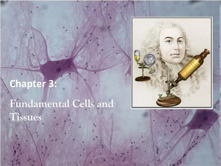
Chapter 3: Fundamental Cells and Tissues. Cells. In the late 1600’s British born Robert Hooke coined the term "cell" to describe the basic unit of life. Where did the term cell come from?

E N D
Chapter 3: Fundamental Cells and Tissues
Cells In the late 1600’s British born Robert Hooke coined the term "cell" to describe the basic unit of life. • Where did the term cell come from? • In 1665 Robert Hooke inspected thin slices of cork and found that they consisted of millions of small, “irregular units” • He used the term cell because the small, bare spaces reminded him of cells in a prison
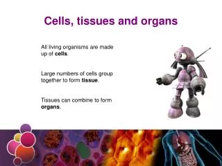
Cell Basics • Cells are the building blocks of all plants and animals • All cells come from the division of preexisting cells • Cells are the smallest units that perform all vital physiological functions • Each cell maintains homeostasis at the cellular level
Cell Basics continued Living cells are about 60 % H20 All body cells are constantly bathed in a dilute saltwater solution called interstitial fluid (18 pints). All exchanges between cells and blood are made through this fluid
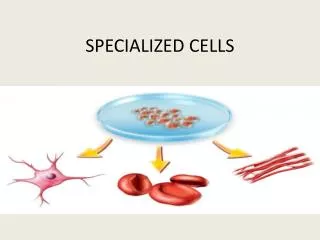
Cell Facts • The human body contains trillions of cells (200 types of cells) • The average cells is just 0.02mm across • The largest cell in the body is an egg cell (0.15mm) • The longest cells are the nerve cells supplying your legs (some measure as much as 4ft in length) • Five million of your body cells die every second
Tissues Tissues - Groups of cells similar in structure and function • The four types of tissues: • Epithelial • Connective • Muscle • Nerve (Neural)
Epithelial Tissue Epithelium – A sheet of cells that covers a body surface or lines a body cavity. Occurs in the body in two ways: 1. Epithelia – covering and lining epithelium - Forms the outer layer of the skin - Lines the open cavities of the cardiovascular, digestive, urinary and respiratory systems and covers the walls and organs 2.Glandular Epithelium - Forms the glands of the body
Special Characteristics of Epithelium • Cells fit closely together to form continuous sheets (single/multiple layers) • Membranes always have one free surface or edge (exposed to body cavity, exterior of the body or lining an internal organ) • Lowest surface (basal surface) of epithelium rests on a basement membrane • Epithelial tissues have no blood supply (avascular) of their own; comes from nearby tissues by way of diffusion • If well nourished, epithelial cells regenerate themselves (high rate of mitosis)
Epithelial Tissue Epithelial Functions include: • Protection – against injury, invasion by bacteria, loss of water and heat • Permeability – selective absorption • Sensation – provide information about the external and internal environments (example - sense smell) • Secretion – releases a substance (example – mucus)
Classification of Epithelial Tissue Cell Shape: • Squamous = flat (attach like tiles) • Cuboidal = thicker, cube shaped • Columnar = tall, cylindrical • Transitional = change shape from columnar/cuboidal to flat due to stretching
Classification of Epithelia Squamous(skwa’mus) Flattened and scalelike Cubodial (ku-boi’dahl) Boxlike, as tall as they are wide. Columnar Tall and column shaped. Figure 4.1b
Classification of Epithelial Tissue Number of cell layers: • Simple = one layer (squamous, cuboidal, columnar) • Stratified = Multilayered (squamous, cuboidal, columnar, transitional) • Pseudostratified Columnar= one layer but appears multilayered (not all cells reach the surface)
Classification of Epithelial Tissue Each epithelium is given two names. • The first name indicates the number of cell layers present • The second name describes the shape of its cells • Followed by epithelium or epithelial tissue Example : • Simple (1 layer) • Squamous (Flat cell shape) • Epithelium
Epithelia: Simple Squamous Figure 4.2a
Epithelia: Simple Cuboidal • Single layer of cubelike cells with large, spherical central nuclei • Function in secretion and absorption • Present in kidney tubules, ducts and secretory portions of small glands, and ovary surface Figure 4.2b
Epithelia: Simple Columnar Figure 4.2c
Epithelia: Pseudostratified Columnar • Single layer of cells with different heights; some do not reach the free surface • Nuclei are seen at different layers • Function in secretion and propulsion of mucus • Present in the male sperm-carrying ducts (nonciliated) and trachea (ciliated) Figure 4.2d
Epithelia: Stratified Squamous – Most Common • Thick membrane composed of several layers of cells • Function in protection of underlying areas subjected to abrasion • Forms the external part of the skin’s epidermis (keratinized cells), and linings of the esophagus, mouth, and vagina (nonkeratinized cells) Figure 4.2e
Epithelia: Stratified Cuboidal and Columnar Stratified cuboidal • Quite rare in the body • Found in some sweat and mammary glands • Typically two cell layers thick Stratified columnar • Limited distribution in the body • Found in the pharynx, male urethra, and lining of some glandular ducts • Also occurs at transition areas between two other types of epithelia
Epithelia: Transitional • Several cell layers, basal cells are cuboidal, surface cells are dome shaped • Stretches to permit the distension of the urinary bladder • Lines the urinary bladder, ureters, and part of the urethra Figure 4.2f
Epithelia: Glandular Gland - One or more cells that makes and secretes a particular product (secretion). Classified by: • Site of product release endocrine (internally secreting) exocrine (externally secreting) • Relative number of cells forming the gland unicellular (one-celled) multicellular (many-celled)
Endocrine Glands • Ductless glands that produce hormones • Hormones are regulatory chemicals that enter the blood or lymphatic fluid and travel to organs • Each hormone prompts its target organ to respond in some characteristic way
Exocrine Glands • More numerous than endocrine glands • Secrete their products onto body surfaces (skin) or into body cavities • Examples include mucus, sweat, oil, salivary glands, the liver (secretes bile), the pancreas (synthesizes digestive enzymes) • Unicellular & Multicellular
Mitosis • Equal division of material in the nucleus, followed by division of the cell body (nuclear cell division) • Epithelial cells have a high rate of mitosis to allow for frequent repair