Download
1 / 31
400 likes | 931 Vues
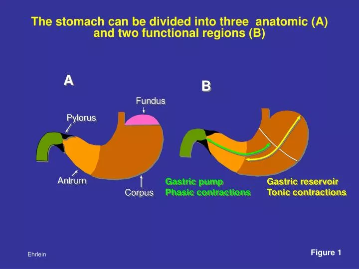
A. B. Fundus. Pylorus. Antrum. Gastric pump Phasic contractions. Corpus. The stomach can be divided into three anatomic (A) and two functional regions (B). Gastric reservoir Tonic contractions. Figure 1. Ehrlein. 1. Receptive relaxation. Mechanical stimuli in the pharynx.

E N D
A B Fundus Pylorus Antrum Gastric pump Phasiccontractions Corpus The stomach can be divided into three anatomic (A) and two functional regions (B) Gastric reservoir Tonic contractions Figure 1 Ehrlein
1. Receptive relaxation Mechanical stimuli in the pharynx Vagus centre Inhibitory vagal fibre (NANC-inhibition) 3. Feedback relaxation 2. Adap tive relax ation ACH NO + VIP et al. CCK Relaxation of gastric reservoir Tension receptors Nutrients Nutrients Distension Figure 2 Ehrlein The relaxation of the gastric reservoir is mainly regulated by reflexes. Three kinds of relaxation can be differentiated: the receptive, adaptive and feedback-relaxation
Tonic contraction Pylorus Accumulation of chyme Peristaltic wave (Pump of the reservoir) Proximal antrum Backflow from antrumand flow from reservoir Figure 3 Ehrlein The transport of digesta from the gastric reservoir into the antral pump is caused by two mechanisms: tonic contractions and peristaltic waves in the region of the gastric corpus
Phases ABC Pylorus Propulsion of chyme into relaxing terminal antrum + duodenal contraction Proximal antrum PA Middle antrum B Phase of emptying Contraction of middle antrum (MA) Terminal antrum Transpyloric and retrograde flow + duodenal relaxation MA closed Pylorus C Phase of retropulsion Contraction of terminal antrum (TA) open TA Duodenum Jet-like back-flow with grinding + duodenal contraction 10 sec Figure 4 Ehrlein The function of the gastric pump can be differentiated into three phases: A: phase of propulsion,B: phase of emptying, C: phase of retropulsion and grinding A Phase of propulsion Contraction of proximal antrum (PA)
Phase of propulsion Phase of emptying Phase of retropulsion Antrum Bulge Emptying of liquids with small particles whereas large particles are retained in the buldge of the terminal antrum Rapid flow of liquids with suspended small particles and delayed flow of large particles towards pylorus Liquids and small particles leave the stomach more rapidly than large particles. This discrimination is called „sieving function“ Retropulsion of large particles and clearing of the terminal antrum Figure 5 Ehrlein
Onset of terminal antral contraction Late phase of terminal antral contraction Pylorus closed Pylorus closing Grinding of solid particles is caused by a forceful jet-like retropulsion through the small orifice of the terminal antral contraction Figure 6 Ehrlein
Phases of gastric emptying Middle antrum Terminal antrum Antral waves closed Pylorus open 9.9 6.6 9.9 3.5 sec 3.5 Proximal duodenum 1 2 1 2 1 4 2 3 3 1 3 Lacking duodenal contractions 0 5 10 15 20 25 30 35 sec Because of different frequencies between antral and duodenal contractions, the duodenum can contract three to four times during an antral wave Figure 7 Ehrlein Antro-duodenal co-ordination: Contractions of the proximal duodenum cease during the phases of gastric emptying..
A. Rapid emptying B. Delayed emptying Pylorus 6a 1a 9 3 8 4 10 6b 1b 5 2 7 Several factors of gastric and duodenal motility co-operate and modulate gastric emptying: A. Rapid emptying is caused by tonic contractions of the reservoir (1a), deep peristaltic waves along the gastric body (1b), deep constrictions of the antral waves (2), a wide opening of the pylorus (3), a duodenal receptive relaxation (4) and peristaltic duodenal contractions (5). B. Delayed emptying due to feedback inhibition is caused by a prolonged relaxation of the reservoir (6a), shallow peristaltic waves along the gastric body ( 6b), shallow antral waves (7), a small pyloric opening (8), a lacking duodenal relaxation (9) and segmenting duodenal contractions (10). Figure 8 Ehrlein
Balance between gastric reservoir and antral pump Gastro-gastric reflexes Enhanced and prolonged relaxation of reservoir Inhibitory reflex Distension Disten- sion Antral pump switched on and intensified Excitatory reflex Figure 9 Ehrlein
Pyloric activity is modulated by antral inhibitoryand duodenal excitatory reflexes Descending inhibitory reflex causing pyloric relaxation Ascending excitatory reflex causing pyloric contractions and increasing pyloric tone Contraction of middle antrum Duodenal stimuli Figure 10 Ehrlein
Pyloric closure Antrum closed Pylorus open Inhibition Duod. bulb Duodenum Stimulation 0.5 ml oleic acid + bile into duodenum An additional function of the pyloric sphincter is to prevent duodeno-gastric reflux Duodenal stimuli like oleic acid inhibit antral contractions, evoke duodenal contractions, increase pyloric tone and elicit frequent pyloric contractions Figure 11 Ehrlein
Lag phase 100 Solids 80 Viscous content 60 Gastric volume (%) 40 Liquid content 20 0 0 80 40 60 20 100 120 Time (min) Solids and liquids of the gastric chyme are emptied with different velocities. Emptying ofliquids isexponential,emptying of largesolid particlesonly begins after sufficientgrinding(lag phase). Afterwards the viscous chyme is mainly emptied in a linear fashion Figure 12 Ehrlein
Non-caloric meal Nutrient meal Feedback control causes Reduced force of antral contractions Antrum closed Reduced pyloric opening Pylorus open Reduced peristaltic waves Duodenal bulb Enhanced segmenting activity Middle Duodenum Nutrients in the gut activate a feedback control and modulate gastric and duodenal motility Gastrointestinal motor patterns after a non-caloric and a nutrient meal Figure 13 Ehrlein
Vagal center Sensoric afferent fibers + Nutrients Long chain fatty acids Amino acids Dipeptids Glucose Osmolality Hydrochloric acid _ + Inhibitory vagal fibers Stimulating cholinergic vagal fibers ACH NO, VIP et al. CCK Enhanced relaxation and storage ACH Reduced opening of pyloric sphincter Backflow Reduced contraction The feedback regulation of gastric emptying is performed by entero-gastric reflexes and release of intestinal hormones It causes enhanced relaxation of the gastric reservoir, inhibition of the antral pump, and reduced opening of the pyloric sphincter. Figure 14 Figure 14 Ehrlein Ehrlein
oral aboral 1 minute 1 minute 1 minute Contractile patterns of the small intestine Peristaltic Stationary Clusters wavescontractionsof contractions The most frequent patterns are peristaltic waves (dashed lines),stationary contractions(arrows), andclusters of contractions, which occur either stationaryat an intestinal segmentor slowlymigrate aborally Figure 15 Ehrlein
Jejunal phase III (MMC) 1 minute oral Velocity of the peristaltic waves Aboral migration of phase III aboral Phase III of the interdigestive motility designated as ”migrating motor complex” (MMC) Rectangles: strain gauge transducers, Data of dog. Figure 16 Ehrlein
Aboral giant contractions Antiperistaltic waves oral oral 0,2 Newton Duodenum Jejunum aboral aboral 1 minute 1 minute Pathological contractile patterns of the proximal intestine Alternating peristaltic (blue arrows) and antiperistaltic waves (red arrows). Giant contractions sometimes originate as a cluster. Figure 17 Ehrlein
Stationary Single segmenting contractions peristaltic waves Excitation Excitation PP 1 1 PP 2 2 3 3 Different kinds of contractile patterns are caused by different kinds of excitation Time course Stationary segmenting contractions are produced by brief excitation of a short intestinal segment Single peristaltic waves are produced by short excitations of a long intestinal segment 1, 2, 3 successive pacesetter potentials (PP) Figure 18 Ehrlein
Stationary cluster Migrating cluster 1 1 2 2 3 3 Time course Stationary excitation Aboral migrating excitation Origin of clustered contractions Clustered contractions are produced by a long lasting excitation of a short intestinal segment. The cluster is stationary when the excitation remains at the same segment. When the excitation slowly moves aborally the cluster of contractions migrates along the intestine. 1, 2, 3 successive pacesetter potentials (PP) Figure 19 Ehrlein
Vagal centre Vago-vagal reflexes Interneurons Integrating circuits Intestinal wall Contractile patterns Sensory neurons Motorneurons Program circuits Enteric nervous system Peptide (CCK) Receptors Intestinal lumenl Glucose - Osmolality Long chain fatty acids Amino acids Central and peripheral control of contractile patterns Luminal stimuli elicit vago-vagal reflexes which activate integrating and program circuits of the enteric nervous system. These activate specific motorneurones responsible for specific contractile patterns. Figure 20 Ehrlein
oral 0,2 Newton aboral Postprandial contractile patterns of the small intestine They are composed of stationary segmenting contractions (green arrows), stationary and migrating clusters of contractions (red horizontal lines) and single short peristaltic waves (dotted lines). Figure 21 Ehrlein
Interdigestive Cycles Phases II III I III Phase III Phase I Phase II Contraction of reservoir Stomach Phase III Sporadic peristaltic waves Pylorus Forceful peristaltic waves Motor quiescence of stomach and duodenum Duodenum Accumulation of residues of chyme Segmenting contractions and single peristaltic waves Jejunum Aboral migration Phase II Motor quiescence Ileum Phase I Phase III The interdigestive motility consists of three phases The phase III of the migrating motor complex originates simultaneously at the stomach and duodenum and migrates within 90 to 120 minutes along the small intestine (dog) Figure 22 Ehrlein
Gastric phase III consisting of 1 - 3 forceful contractions of the gastric reservoir and lumen occluding peristaltic waves occurring at intervals of 2-3 min Gastric phases III Middle Antrum 0 mm Pyloric diameter 6 mm Duodenal bulb Duodenum 1 min Stomach is cleaned of residues of chyme and secretions. A P P P The antral waves are associated with a wide opening of the pylorus and inhibition of duodenal contractions followed by duodenal peristaltic waves occurring at maximal frequency. Figure 23 Ehrlein
Intestinal phase III Time (about 20 sec) oral Successsive peristaltic waves Chyme Slow aboral migration of phase III aboral Phase III (MMC) of the small intestine The peristaltic waves clean the intestinal segment from chyme which accumulates aborally. Because the successive waves start and end further aborally the phase III slowly migrates distally Figure 24 Ehrlein
Fed motor pattern Phase III Meal Antrum closed Pylorus open Duodenum 5 min Ingestion of a meal suppresses the interdigestive motility and induces a fed motor pattern Postprandial motility is characterised by a lower amplitude of the antral waves occurring at maximal frequency, rhythmic pyloric opening and closure and co-ordinated duodenal contractions occurring in sequence with the antral waves Figure 25 Ehrlein
Shallow peristaltic waves of caecum and colon A low propulsion backflow Shallow peristaltic waves at haustrated colon B small aboral flow Colonic segmenting contractions migrating aborally slow aboral propulsion C aboral migration Contractile patterns of the large intestine C: A special feature of the large intestine are multiple segmenting contractions of long duration migrating aborally. They divide digesta into boli pushing them slowly aborally. The motility tracings show a rise of the baseline superimposed by phasic contractions Figure 26 Ehrlein
Motility of the large intestine in pig Ileum - Caecum - Colon A B Caecum J1 C1 Giant contractions J2 C2 C1 Colonic wave C3 Co1 1 min Co2 Co3 Colon Co3 Co2 Ileum 1 min J1 A: Haustral movements of the caecum result in clustered contractions. B: The ileum is emptied by giant contractions. They occur either isolated or in co-ordination with peristaltic waves of the caecum and colon. Additional colonic waves originate at the beginning of the colonic coil. Co1 J2 Caecum Distal colon C1 C2 C3 Ehrlein Figure 27
Caecum Colon Spiral colon Giant contraction C4 Peristaltic wave C1 C3 SC1 Co1 C2 SC2 C1 Co2 SC3 Co1 Co3 Ileum C3 C4 C2 Co2 Co1 Co4 C1 Colon 1 min Caecum 1 min Co2 Co3 Co2 Caecal motility is characterised by peristaltic and antiperistaltic waves. In the colon peristaltic waves and giant contractions are the dominant feature. In the spiral colon prolonged segmenting contractions divide digesta into boli and push them distally. SC1 SC2 Co4 SC3 Spiral colon Figure 28 Ehrlein Motility of caecum and colon in sheep.
Colon Giant contractions J1 Co1 C1 Co2 C2 Co3 1 min C3 Co3 Co2 J1 Ileum Co1 C4 C1 Colon C2 C5 Caecum 1 min C5 C4 C3 Motor patterns of the large intestine in rabbits Caecum Caecal motility is characterised by peristaltic and antiperistaltic waves. Migrating segmenting contractions are the dominant feature of the single haustrated colon. Figure 29 Ehrlein
A B Colonic motor complex (CMC) oral Phasic contractions 5 min 15 min aboral Colonic motor complexes (CMC’s) of the canine colon A: Slow paper speed. The CMC’s occur at all parts of the colon at intervals of 20-30 min. B: High paper speed. The CMC’s consist of a rise of the baseline super- imposed of phasic contractions. The onset of the CMC‘s obviously differs along the colon (indicated by lines). Figure 30 Ehrlein
Retrograde giant contraction Vomiting Distal duod. Prox. duod. Bulbus closed Pylorus (P) open Antrum 1 3 2 5 4 1 min Duodenum P P P Jejunum Stomach 3 1 5 4 2 Retrograde giant contraction followed by vomiting (1) Normal segmenting contractions of the proximal jejunum (2) Start of a retrograde giant contraction in proximal jejunum; (3) Retropelled digesta reach the duodenum and (4) are forced across the widely opened pylorus into the antrum; (5) The giant contraction proceeds to the antrum, the chyme accumulates in the gastric reservoir. Figure 31 Ehrlein