Download
1 / 15
150 likes | 369 Vues
Medical Imaging. By Anuja Kulkarni 1000722132. LIST OF ACRONYMS. CAT - Computed Axial Tomography CT - Computed Tomography DICOM - Digital Imaging and Communications in Medicine EEG - Electroencephalography EKG - Electrocardiography fMRI - Functional Magnetic Resonance Imaging

E N D
Medical Imaging By AnujaKulkarni 1000722132
LIST OF ACRONYMS • CAT - Computed Axial Tomography • CT - Computed Tomography • DICOM - Digital Imaging and Communications in Medicine • EEG - Electroencephalography • EKG - Electrocardiography • fMRI- Functional Magnetic Resonance Imaging • JFIF- JPEG File Interchange Format • JPEG- Joint Photographic Experts Group • JPIP- JPEG 2000 Interactive Protocol • MEG - Magnetoencephalography • MRI - Magnetic Resonance Imaging • NMR- Nuclear Magnetic Resonance • PNG- Portable Network Graphics • RF- Radio Frequency • SSIM- Structural Similarity
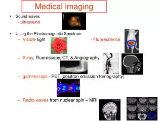
Introduction • Medical imaging as the name suggests is the technique and process used to create images of parts and functions of human body for clinical purposes. • It is a medical procedure seeking to reveal, diagnose or examine disease. [1] There are two types of medical imaging, they are- • Invisible light medical imaging- radiology /clinical imaging • Visible light medical imaging- involves digital video or still pictures that can be seen without special equipment.
Imaging Technologies • Radiology: [2] Two forms of radiographic images are in use in medical imaging; projection radiography and fluoroscopy. Figure 1: Digital Radiography
Magnetic Resonance imaging and fiduciary markers • A magnetic resonance imaging instrument, uses powerful magnets to polarise and excite hydrogen nuclei in water molecules in human tissue, producing a detectable signal which is spatially encoded, resulting in images of the body. [3] • Fiduciary Markers Fiduciary markers are used in a wide range of medical imaging applications. Images of the same subject produced with two different imaging systems may be correlated by placing a fiduciary marker in the area imaged by both systems. [4] Figure 2: fMRI scan Figure 3: Fiducial Marker Example
Photo-acoustic imaging and tomography • Photo acoustic imaging is a recently developed hybrid biomedical imaging modality based on the photo acoustic effect. [5] • Tomography is the method of imaging a single plane, or slice, of an object resulting in a tomogram. [5] • Figure 4: Computed tomography of brain
Creation of three-dimensional images • Recently, techniques have been developed to enable CT, MRI and ultrasound scanning software to produce 3D images for the physician. [6] • To produce 3D images, many scans are made, then combined by computers to produce a 3D model, which can then be manipulated by the physician. • Other proposed or developed techniques include: • Diffuse optical tomography • Elastography • Electrical impedance tomography • Optoacoustic imaging • Ophthalmology
Compression of medical images • JPEG 2000 is the state-of-the-art image compression DICOM standard for storage and transmission of medical images. [7] Figure 5: Comparison of JPEG2000 with JPEG [8]
Compression of medical images • JPIP (JPEG 2000 Interactive Protocol) is a compression streamlining protocol that works with JPEG 2000 to produce an image using the least bandwidth required. [9] • JPIP has the capacity to download only the requested part of a picture, saving bandwidth, computer processing on both ends, and time. [10]
BLOCK DIAGRAM OF JPEG 2000 [11] Figure 6: Block diagram of JPEG Encoder/decoder [11]
Non-diagnostic imaging (Neuroimaging) • Neuroimaging has also been used in experimental circumstances to allow people to control outside devices, acting as a brain computer interface. [12] • Neuroimaging falls into two broad categories: • Structural imaging • Functional imaging which is used to diagnose metabolic diseases and lesions on a finer scale (such as Alzheimer's disease) Figure 7: 3D MRI section of the head [13]
Proposed work • This project introduces the concept of medical imaging and divulges into its technologies like MRI, tomography, ultrasound etc. It will also compare the compression techniques of medical imaging i.e. JPEG2000 and JPIP on the basis of their bit rates, SSIM index [14], and complexity. • This project proposes to demonstrate creation of 3D images of CT/MRI scan from a normal 2D image. It also shows some circumstances of neuroimagingi.e non-diagnostic medical imaging as in Figure 6.
References • http://en.wikipedia.org/wiki/Medical_imaging • L.F.Squire and R.A.NovellineSquire's fundamentals of radiology (5th ed.). Harvard University Press. ISBN 0-674-83339-2 1997; http://en.wikipedia.org/wiki/Image:FMRI.jpg • M. Xu and L.H. Wang ; "Photoacoustic imaging in biomedicine". Review of Scientific Instruments77 (4): 041101. doi:10.1063/1.2195024; 2006 • G.T.Herman, “Fundamentals of computerized tomography: Image reconstruction from projection” , 2nd edition, Springer, 2009 • S.Richard., and C. Cobbold, Foundations of Biomedical Ultrasound, Oxford University press pp. 422–423. 978-0-19-516831-0 • A. Yamani; A novel pulse-echo technique for medical three dimensional imaging; IEEE trans med imaging; Volume 16 Issue 6 Pages 938-942; Dec 1997 • A.Khademi and S.Krishnan ; Comparison of JPEG 2000 and other lossless compression schemes; Paper in Engineering in medicine and biology society (IEEE EMBS);2005
References 8.http://en.wikipedia.org/wiki/File:JPEG_JFIF_and_2000_Comparison.png(Figure 5) 9. eeweb.poly.edu/~yao/EE3414/JPEG.pdf (figure 6) 10.http://en.wikipedia.org/wiki/File:JPEG_JFIF_and_2000_Comparison.png (Figure 6) 11. Microsoft and NASA Bring Mars Down to Earth Through the WorldWide Telescope (07.12.10) - NASA 12. A.G.Filler, “The history, development, and impact of computed imaging in neurological diagnosis and neurosurgery: CT, MRI, DTI: Nature Proceeding DOI: 10.1038/npre.2009.3267.5.Neurosurgical Focus (in press); 2009 13. http://en.wikipedia.org/wiki/Neuroimaging (Figure7)/ Picture reference: sbharris on wikipedia 14. http://en.wikipedia.org/wiki/Structural_similarity