Download
1 / 28
280 likes | 300 Vues
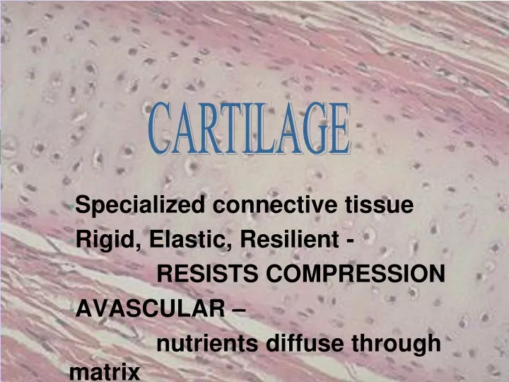
CARTILAGE. Specialized connective tissue Rigid, Elastic, Resilient - RESISTS COMPRESSION AVASCULAR – nutrients diffuse through matrix. Hundreds of Eyes Staring Back at YOU!. PERICHONDRIUM. Dense irregularly arranged connective tissue (type I collagen)

E N D
CARTILAGE Specialized connective tissue Rigid, Elastic, Resilient - RESISTS COMPRESSION AVASCULAR – nutrients diffuse through matrix
PERICHONDRIUM • Dense irregularly arranged connective tissue (type I collagen) • Ensheaths the cartilage • Houses the blood vessels that nourish chondrocytes
CHONDROBLAST • Progenitor of chondrocytes • Lines border between perichondrium and matrix • Secretes type II collagen and other ECM components • Chondroblasts build
CHONDROCYTE • Mature cartilage cell • Reside in a space called the lacuna • Clear areas = Golgi and lipid droplets
Chondrocytes completely fill their lacunae • RER and euchromatic nuclei • Synthetically active, secrete matrix N RER Cartilage matrix
MATRIX • Provides the rigidity, elasticity, & resilience • FIBERS • Collagenous and elastic • GROUND SUBSTANCE • Glycosaminoglycans (chondroitin sulfates, keratin sulfate, hyaluronic acid) • Proteoglycans: GAGs + core protein • Water • Basophilic • Territorial matrix - high [ ] of sulfated proteoglycans
CARTILAGE GROWTH • Appositional • Increasing in WIDTH; chondroblasts deposit matrix on surface of pre-existing cartilage • Interstitial • Increasing in LENGTH; chondrocytes divide and secrete matrix from w/in lacunae
Cartilage • Embryo • More prevalent than in adult • Skeleton initially mostly cartilage • Bone replaces cartilage in fetal and childhood periods
Location of cartilage in adults • External ear • Nose • “Articular” – covering the ends of most bones and movable joints • “Costal” – connecting ribs to sternum • Larynx - voice box
Epiglottis – flap keeping food out of lungs • Cartilaginous rings holding open the air tubes of the respiratory system (trachea and bronchi) • Intervertebral discs • Pubic symphysis • Articular discs such as meniscus in knee joint
Remember the four basic types of tissue… • Epithelium • Connective tissue • Connective tissue proper • Cartilage • Bone • Blood • Muscle tissue • Nervous tissue
Cartilage is connective tissue • Cells called chondrocytes • Abundant extracellular matrix • Fibers: collagen & elastin • Jellylike ground substance of complex sugar molecules • 60-80% water (responsible for the resilience) • No nerves or vessels (hyaline cartilage)
TYPES OF CARTILAGE • HYALINE • ELASTIC • FIBROUS
Types of cartilage: 3 • Hyaline cartilage: flexible and resilient • Chondrocytes appear spherical • Lacuna – cavity in matrix holding chondrocyte • Collagen the only fiber • Elastic cartilage: highly bendable • Matrix with elastic as well as collagen fibers • Epiglottis, larynx and outer ear • Fibrocartilage: resists compression and tension • Rows of thick collagen fibers alternating with rows of chondrocytes (in matrix) • Knee menisci and annunulus fibrosis of intervertebral discs
HYALINE CARTILAGE • FUNCTION • Support tissue and organs • Model for bone development • MATRIX • Type II collagen (thin fibrils) • Chondroitin sulfate, keratin sulfate, hyaluronic acid • Water • LOCATION • Tracheal rings, nasal septum, larynx, articular surfaces of joints
ELASTIC CARTILAGE • FUNCTION • Support with flexibility • MATRIX • Normal components of hyaline matrix plus ELASTIC fibers • LOCATION • External ear, external auditory canal, epiglottis • STAINS • Elastic fibers stain BLACK with Weigert stain perichondrium
FIBROCARTILAGE • FUNCTION • Support with great tensile strength • MATRIX • Type I collagen - Oriented parallel to stress plane • LOCATION • Intervertebral disks, pubic symphysis
FIBROCARTILAGE • Chondrocytes align between collagen fibers • Collagen fibers lie parallel to lines of stress
Before we look at collagen pic… • Hyaline cartilage: flexible and resilient • Chondrocytes appear spherical • Lacuna – cavity in matrix holding chondrocyte • Collagen the only fiber • Elastic cartilage: highly bendable • Matrix with elastic as well as collagen fibers • Epiglottis and larynx • Fibrocartilage: resists compression and tension • Rows of thick collagen fibers alternating with rows of chondrocytes (in matrix) • Knee menisci and annulus fibrosis of intervertebral discs
Triple helix of collagen molecules form fibril Fibrils aggregate into collagen fibers
Growth of cartilage • Appositional • “Growth from outside” • Chrondroblasts in perichondrium (external covering of cartilage) secrete matrix • Interstitial • “Growth from within” • Chondrocytes within divide and secrete new matrix • Cartilage stops growing in late teens (chrondrocytes stop dividing) • Regenerates poorly in adults
Uploaded By.... M.Farrukh Fayyaz