Download
1 / 37
380 likes | 580 Vues
Immunoglobulins. Biological Properties. Introduction . Many important biological properties are attributed to antibodies that differ depending on isotype These include; - Neutralization of toxins - Immobilization of microorganisms

E N D
Immunoglobulins Biological Properties
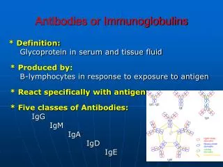
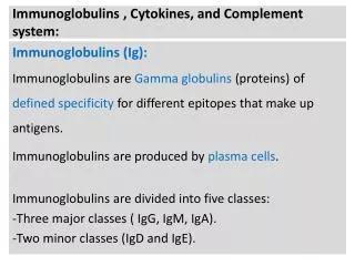
Introduction • Many important biological properties are attributed to antibodies that differ depending on isotype • These include; - Neutralization of toxins - Immobilization of microorganisms - Neutralization of viral infectivity - Agglutination of microorganisms or antigenic particles - Binding with soluble antigens - Activation of complement - Protection of fetus
Immunoglobulin Structure-Function Relationship • Cell surface antigen receptor on B cells • - Allows B cells to sense their antigenic environment • - Connects extracellular space with intracellular • signalling machinery • Secreted antibody • - Neutralization • - Arming/recruiting effector cells • - Complement fixation
Immunoglobulins are Bifunctional Proteins • Immunoglobulins must interact with a • small number of specialized molecules : • - Fc receptors on cells • - Complement proteins • - Intracellular cell signalling molecules • Whilst simultaneously recognising an infinite • array of antigenic determinants.
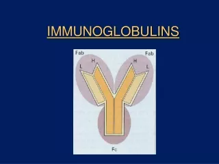
Why do antibodies need an Fc region? • The (Fab)2 fragment can – • Detect antigen • Precipitate antigen • Block the active sites of toxins or pathogen- associated molecules • Block interactions between host and pathogen- associated molecules
but the (Fab)2 can not activate • Inflammatory and effector functions associated with cells • Inflammatory and effector functions of complement • The trafficking of antigens into the antigen processing pathways
Four distinct roles of Fc binding proteins • They are essential for many of the biological functions of antibodies: 1- The movement of Ab across cell membranes : poly IgR for dimericIgA & to some extent, pentamericIgM 2- The transfer of lgG from mother to fetus across the placenta : FcRN 3- Trigger effector functions : Opsonization or ADCC 4- Cross-linking of FcR which generates immunoregulatory signals that affect cell activation, differentiation, etc. which are similar to signal transduction from BcR
The structure of a number of human Fc -receptors Fc -binding polypeptide Accessory signal- transducing polypeptide
Biological Properties of IgG • Distributed equally between the intravascular and extravascular spaces • Except for IgG3 which has a rapid turnover (half life=7days), the half life of IgG is approximately 23 days • IgG has the longest half life of all immunoglobulin isotypes making it the most suitable for passive immunization • Interestingly, as the concentration of IgG in the serum increases, the rate of IgG catabolism increases (half life 15-20 days)
Functions of IgG • Agglutination and precipitation • Passage through placenta - The IgGisotype, except for IgG2, is the only isotype that can pass through the placenta as of the 3rd to 4th month of gestation - Passage is mediated by the FC portion - Role in health and disease • Opsonization - Bridges microorganisms or particulate antigens to phagocytic cells
ADCC - NK cells • Activation of Complement -Classical or alternative pathway • Neutralization of toxins - Excellent function against toxins such as tetanus and botulinum toxins - Inactivation of snake or scorpion venoms by blocking the active site
Immobilization of Bacteria - IgG molecules are efficient in immobilizing bacteria - Reaction of IgG specific to flagella cause organisms to clump arresting their movement • Neutralization of Viruses -IgG is an efficient virus neutralizing antibody - Act by inhibiting attachment, penetration, uncoating, or later steps
S S S S s s C J C S S C C S S C C IgA dimerisation and secretion IgA is the major isotype of antibody secreted at mucosal surfaces Exists in serum as a monomer, or as a J chain-linked dimer, that is formed in a similar manner to IgM pentamers. IgA exists in two subclasses IgA1 is mostly found in serum and made by bone marrow B cells IgA2 is mostly found in mucosal secretions, colostrum and milk and is made by B cells located in the mucosa
S S S S S S S S S S S S S S S S S S S S IgA and pIgR are transported to the apical surface in vesicles Epithelial cell s s s s s s s s s s C C C C C J J J J J C C C C C S S S S S S S S S S C C C C C C C C C C pIgR & IgA are internalised S S S S S S S S S S Polymeric Ig receptors are expressed on the basolateral surface of epithelial cells to capture IgA produced in the mucosa C C C C C C C C C C B B cells located in the submucosa produce dimeric IgA Secretory IgA and transcytosis ‘Stalk’ of the pIgR is degraded to release IgA containing part of the pIgR - the secretory component
Properties of IgA • Serum IgA: Half life of 5.5 days, has no important biologic functions • SecretoryIgA: - Important primary immunologic defense against local infections on mucosal surfaces - No complement activity, therefore, no bacterial lysis - Bactericidal for Gram negative bacteria in the presence of lysozyme - Antiviral activity - Agglutinating activity
IgA facts and figures Heavy chains:a1or a2 - Alpha 1 or 2 Half-life: IgA1 5 - 7 days IgA2 4 - 6 days Serum levels (mgml-1): IgA1 1.4 - 4.2 IgA2 0.2 - 0.5 % of Ig in serum: IgA1 11 - 14 IgA2 1 - 4 Complement activation:IgA1 - by alternative and lectin pathway IgA2 - No Interactions with cells: Epithelial cells by pIgR Phagocytes by IgA receptor Transplacental transfer: No To reduce vulnerability to microbial proteases the hinge region of IgA2 is truncated. In IgA1 the hinge is heavily glycosylated. IgA is inefficient at causing inflammation and elicits protection by excluding, binding, cross-linking microorganisms and facilitating phagocytosis
Biologic Properties of IgM • Predominantly found in the intravascular space • Half life is about 5 days • It is the only immunoglobulin class synthesized by the fetus beginning at approximately 5 months of gestation • It is the first antibody to be produced and its presence indicates a recent infection
Functions of IgM • Agglutination -Very efficient - Forms bridges between distant antigenic epitopes • Isohemagglutinins - Naturally occurring against RBC antigens - Triggered by exposure to bacteria bearing similar determinants - Transfusion reactions • Activation of Complement - Most efficient complement fixing antibody
Cm4 Cm2 Cm3 Cm1 Monomeric IgM IgM only exists as a monomer on the surface of B cells Monomeric IgM has a very low affinity for antigen Cm4 contains the transmembrane and cytoplasmic regions. These are removed by RNA splicing to produce secreted IgM
Cm4 Cm2 Cm3 Cm1 Polymeric IgM IgM forms pentamers and rarely hexamers Cm3 binds C1q to initiate activation of the classical complement pathway Cm1 binds C3b to facilitate uptake of opsonised antigens by macrophages Cm4 mediates multimerisation (Cm3 may also be involved)
Multimerisation of IgM C C Cm4 Cm4 Cm4 Cm4 Cm4 Cm2 Cm2 Cm2 Cm2 Cm2 Cm3 Cm3 Cm3 Cm3 Cm3 C s s s s s s s s C C C C C C C C C C C C C C C 1. Two IgM monomers in the ER (Fc regions only shown) 2. Cysteines in the J chain form disulphide bonds with cysteines from each monomer to form a dimer 3.A J chain detaches leaving the dimer disulphide bonded. 4.A J chain captures another IgM monomer and joins it to the dimer. 5. The cycle is repeated twice more 6. The J chain remains attached to the IgM pentamer.
IgM facts and figures Heavy chain: m - Mu Half-life: 5 to 10 days % of Ig in serum: 10 Serum level (mgml-1): 0.25 - 3.1 Complement activation:++++ by classical pathway Interactions with cells: Phagocytes via C3b receptors Epithelial cells via polymeric Ig receptor Transplacental transfer: No Affinity for antigen: Monomeric IgM - low affinity - valency of 2 Pentameric IgM - high avidity - valency of 10
Biological Properties of IGD &IgE • IgD - No function except B cell maturation - Half life is 2-8 days • IgE ( Reaginic antibody) -Half life is 2 days - Binds with high affinity to mast cells and basophils -No agglutination or complement fixing activities - Antiparasitic - Major role in hypersensitivity
IgD facts and figures Heavy chain: d - Delta Half-life: 2 to 8 days % of Ig in serum: 0.2 Serum level (mgml-1): 0.03 - 0.4 Complement activation:No Interactions with cells:T cells via lectin like IgD receptor Transplacental transfer: No IgD is co-expressed with IgM on B cells due to differential RNA splicing Level of expression exceeds IgM on naïve B cells IgD plasma cells are found in the nasal mucosa - however the function of IgD in host defence is unknown Ligation of IgD with antigen can activate, delete or anergise B cells
IgE facts and figures Heavy chain: e - Epsilon Half-life: 1 - 5 days Serum level (mgml-1):0.0001 - 0.0002 % of Ig in serum: 0.004 Complement activation:No Interactions with cells:Via high affinity IgE receptors expressed by mast cells, eosinophils, basophils and Langerhans cells Via low affinity IgE receptor on B cells and monocytes Transplacental transfer: No IgE appears late in evolution in accordance with its role in protecting against parasitic infections Most IgE is absorbed onto the high affinity IgE receptors of effector cells IgE is also closely linked with allergic diseases
antigen Role for IgE on mast cells and basophils High affinity receptor for IgE antigen Antigen comes to the mast cell which already has IgE attached to its receptor
Passive Sero - & Antibody therapy • ① In 1890, injection of 0.2ml serum from tetanus-immunized rabbits into the abdominal cavity of mice protected from challenge of virulent tetanus bacteria (Dr. Von Behring) • During the 1930s & 1940s, passive immunotherapy based on the transfer of Ab (measles & Hepatitis A) was used in clinical (medical) practice.
Sero -therapy Tetanus toxoid Immunized horse Immune horse serum (tetanus antitoxin) Patient at risk of tetanus Patient protected The passive transfer of immunity to tetanus by means of antibody
Passive Immunity • Immune protection produced by transfer of antibodies to a recipient from a donor • Donor has been actively immunized • Occurs naturally from mother to fetus during pregnancy and mother to infant during nursing • Short-lived protection
Antibody therapy • Pooled plasma from thousands of donors -> treatment with solvents & the use of detergents that was highly effective in inactivating viruses. • Intravenous immune globulin (IVIG) contains ~1018 Ab (mostly IgG) which may incorporate > 107 different Ab specificities • Action mechanism of passively administered Ab. i) Activation of the complement pathway ii) Promotes opsonization, phagocytosis & killing of bacteria iii) mediate the killing of target cells by NK cells (ADCC) iv) neutralizes toxins & viruses
The conventional polyclonal antiserum contains a mixture of monoclonal antibodies
Uses of Monoclonal Antibodies • Diagnostic agents (histology, immunoassays) • Experimental probes for cell biology • Therapeutic agents • What are the advantages over polyclonal antibodies raised by immunisation of larger animals?
CD52 is strongly expressed on lymphocytes and not on blood stem cells CD52+ lymphocytes STEM CELLS MYELOID CELLS PLATELETS RED CELLS