Download
1 / 58
590 likes | 691 Vues
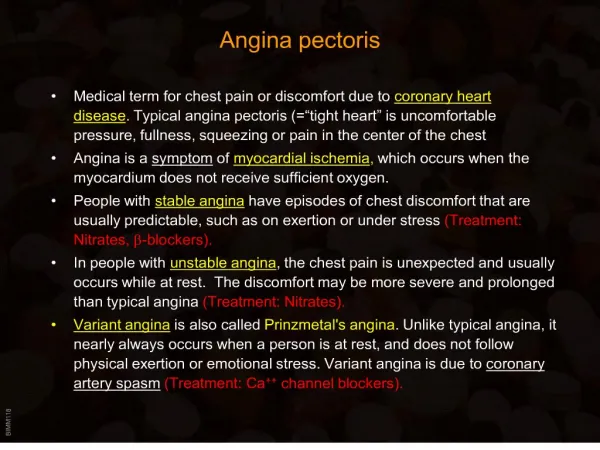
MANAGING ANGINA. JIM McLENACHAN , CONSULTANT CARDIOLOGIST, LEEDS. 17 th November 2011. Myocardial Supply (blood flow). Myocardial Demand (work). Myocardial Ischaemia. What is angina ?. “not a pain” tightness, pressure, heaviness usually in centre of chest

E N D
MANAGING ANGINA JIM McLENACHAN, CONSULTANT CARDIOLOGIST, LEEDS. 17th November 2011
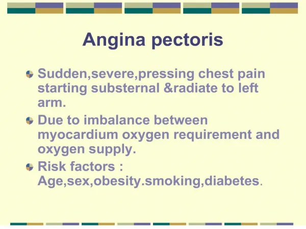
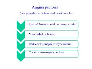
Myocardial Supply (blood flow) Myocardial Demand (work) Myocardial Ischaemia
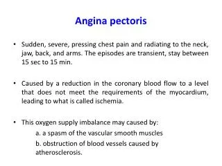
What is angina ? • “not a pain” • tightness, pressure, heaviness • usually in centre of chest • may radiate to either arm, neck, jaw • usually provoked by exercise (walking) • usually relieved by rest • lasts no more than 2 minutes
Differential diagnosis • Dyspepsia • Musculoskeletal pain • Undiagnosed !!
Baseline investigation of suspected angina: • Examination – HR, BP, murmurs • ECG • FBC • U and E • Cholesterol • Glucose
Resting ECG • Limited value • Useful if evidence of old MI • Normal ECG does not exclude extensive coronary disease
NICE Clinical guideline 95(published March 2010) • Estimate risk according to: - non-anginal, atypical, typical angina - age - sex - low or high risk
NICE definition of angina • constricting discomfort in the front of the chest, or in the neck, shoulders, jaw, or arms • precipitated by physical exertion • relieved by rest or GTN within about 5 minutes. 3 out of 3 = typical angina 2 out of 3 = atypical angina 0/1 out of 3 = non-anginal pain
Table 1 Percentage of people estimated to have coronary artery disease according to typicality of symptoms, age, sex and risk factors NICE Guidance CG95Chest pain of recent onsetEstimation of risk
NICE Clinical guideline 95(published March 2010) • Risk <10% - no tests (!) • Risk 10-29% - Cardiac CT scanning • Risk 30-60% - Functional imaging • Risk > 60% - Coronary angiography
Coronary CT scanning • Some technical difficulties • Beta blockers needed to slow heart rate • Very sensitive test • Negative result is useful • Positive result needs more tests!
Functional imaging • Stress myocardial perfusion scanning • Stress echo • Stress MR imaging
Interpretation of Myocardial Perfusion Studies Stress Rest Interpretation Image Image Normal Fixed defect (infarction) Reversible defect (ischaemia)
Angiography in stable angina • Diagnostic doubt • Ischaemia at low workload • Young patients • Ongoing symptoms • Threatened employment
DAY CASE CORONARY ANGIOGRAPHY • Performed under local anaesthetic • Duration 20 – 30 minutes • Arterial access via femoral, brachial or radial artery. • Complications rare: stroke / MI - < 1 in 1,000 haematoma - 5 – 10 %
IS ANGIOGRAPHY THE GOLD STANDARD? • If uncertainty persists, - intravascular ultrasound - pressure wire assessment
Initial treatment of suspected angina: • Aspirin 75 mg once daily • GTN spray (with advice) • Beta blocker (eg. bisoprolol 5mg once daily) • Statin
Treatment of Angina • Aspirin • Short acting nitrate • Beta blocker • Statin • ACE inhibitor Underlined classes are for secondary prevention and are likely to be life-long.
NICE Clinical guidelines (CG126) 1st line drugs • Beta blockers eg. Bisoprolol • Cacium channel blockers eg. diltiazem
NICE Clinical guidelines (CG126) 3rd line drugs(after beta blockers and calcium channel blockers) • a long-acting nitrate or • ivabradineor • nicorandilor • ranolazine
Treatment of Stable Angina • Medical treatment • PCI • CABG
CLOPIDOGREL • Use instead of aspirin if genuine aspirin intolerance. • Consider aspirin plus PPI in aspirin-induced dyspepsia. • Give for 12 months following ACS admission • After stenting: 3 months - bare metal stent, elective 12 months – any drug-eluting stent , any ACS
Criticism of cardiologists’ management of angina Exercise test Chest pain OP assessment Coronary angiogram PCI
Criticism of cardiologists’ management of angina Exercise test Chest pain OP assessment Coronary angiogram PCI one cardiologist
Treatment of Stable Angina .....the oculostenotic reflex....... .....to a man with a hammer, everything looks like a nail....
Treatment of Stable AnginaNICE Clinical Guideline 126(published July 2011) • Optimising medical treatment • Demonstration of ischaemia • Importance of MDT discussion
NICE Clinical guideline 126(published July 2011) • The main purpose of revascularisation is to improve the symptoms of stable angina. • CABG and PCI are effective in relieving symptoms. • Repeat revascularisation may be necessary after either CABG or PCI and the rate is lower after CABG. • Stroke is uncommon after either CABG or PCI, and the incidence is similar between the two procedures. • There is a potential survival advantage with CABG for some people with multivessel disease.
Treatment of refractory angina • Is it really angina? • Stellate ganglion blocks • EECP
NICE Clinical guideline 126(published July 2011) Do not offer the following interventions to manage stable angina: • transcutaneous electrical nerve stimulation (TENS) • enhanced external counterpulsation (EECP) • acupuncture
CLASSIFICATION OF ANGINA / MI • STABLE ANGINA • ACUTE CORONARY SYNDROMES -unstable angina -non ST segment elevation MI (NSTEMI) -ST segment elevation MI (STEMI)
Pathophysiology of ACS Unstable angina Non ST elevation MI NON-STEMI Stable angina ST elevation MI STEMI
Myocardial Infarction • More prolonged chest pain • More severe chest pain • More systemic upset (nausea, sweating, etc.)
Non-ST Elevation MI • Chest pain may be new onset, or more readily induced, or more prolonged than “normal” • ECG may show • transient ST elevation • ST segment depression • T-wave inversion • nothing • Diagnosis often based on troponin
Troponin • Very sensitive - normal troponin 12 hours after onset of pain effectively “rules out” ischaemic pain. • Not very specific - also raised in patients with heart failure, renal failure, atrial fibrillation etc.
Non-ST Elevation MI • Aspirin • Clopidogrel (for 12 months) • Heparin (Fondaparinux) • Statin • Beta blockers • Early angiography (within 48 hours) followed by PCI / CABG as appropriate.
Newer antiplatelet agents • Prasugrel • Ticagrelor
TREATMENT OF ST ELEVATION MI • ASPIRIN • BETA BLOCKERES • THROMBOLYSIS - Streptokinase - TPA - Reteplase - Tenectaplase • PCI
Primary PCI vs.ThrombolysisMortality 95% CI 0.73 [0.06,0.86] 95% CI 0.70 [0.58,0.85] Percent Lancet 2003; 361:13-20 PCI Lytic Lytic PCI No SHOCK Patients All Patients
Primary PCI vs. Thrombolysis p<0.0001 p<0.0001 Percent Lancet 2003; 361:13-20 p=0.0004 p<0.0001 Total Stroke Haemorrhagic Stroke Death, reinfarction, stroke Reinfarction
Author: Dr Huon Gray, Consultant Cardiologist, Southampton. 20th October, 2008