Download
1 / 104
1.11k likes | 1.28k Vues
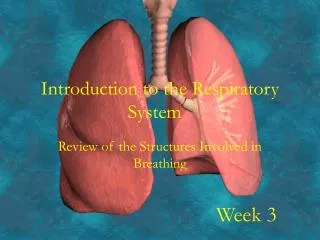
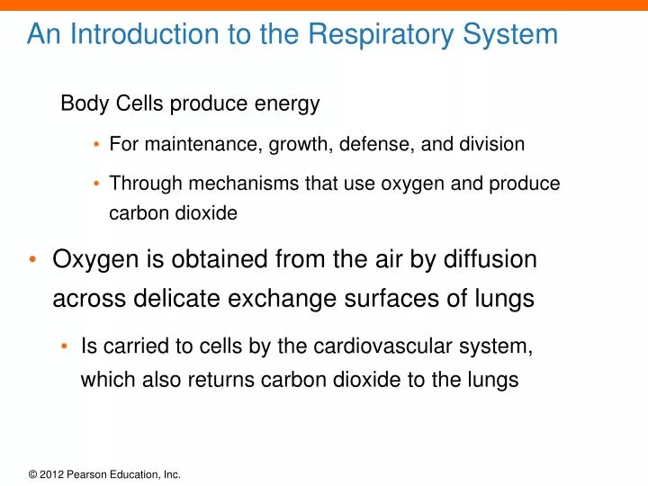
An Introduction to the Respiratory System. Body Cells produce energy For maintenance, growth, defense, and division Through mechanisms that use oxygen and produce carbon dioxide Oxygen is obtained from the air by diffusion across delicate exchange surfaces of lungs

E N D
An Introduction to the Respiratory System Body Cells produce energy • For maintenance, growth, defense, and division • Through mechanisms that use oxygen and produce carbon dioxide • Oxygen is obtained from the air by diffusion across delicate exchange surfaces of lungs • Is carried to cells by the cardiovascular system, which also returns carbon dioxide to the lungs
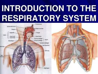
23-1 Components of the Respiratory System • Five Functions of the Respiratory System • Provides extensive gas exchange surface area between air and circulating blood • Moves air to and from exchange surfaces of lungs • Protects respiratory surfaces from outside environment • Produces sounds • Participates in olfactory sense
23-1 Components of the Respiratory System • Organization of the Respiratory System • The respiratory system is divided into: • Upper respiratory system - above the larynx • Lower respiratory system - below the larynx
Figure 23-1 The Components of the Respiratory System RIGHTLUNG LEFTLUNG Frontal sinus Nasal cavity Nasal conchae Sphenoidal sinus Nose Internal nares Tongue UPPERRESPIRATORYSYSTEM Pharynx Hyoid bone Larynx LOWERRESPIRATORYSYSTEM Esophagus Trachea Clavicle Bronchus Bronchioles RIGHTLUNG Ribs Diaphragm
23-5 The Lungs • WHY ARE THE ALVEOLI SO IMPORTANT??? • Respiratory DistressSyndrome • Difficult respiration • Due to alveolar collapse • Caused when pneumocytes type II do not produce enough surfactant • Respiratory Membrane • The thin membrane of alveoli where gas exchange takes place
Figure 23-11c Alveolar Organization Red blood cell Capillary lumen Capillary endothelium Nucleus of endothelial cell 0.5 m Alveolar epithelium Surfactant Fused basement membrane Alveolar air space The respiratory membrane, which consists of an alveolar epithelial cell, a capillary endothelial cell, and their fused basement membranes.
23-5 The Lungs • Diffusion • Across respiratory membrane is very rapid • Because distance is short • Gases (O2 and CO2) are lipid soluble • Inflammation of Lobules • Also called pneumonia • Causes fluid to leak into alveoli • Compromises function of respiratory membrane
23-5 The Lungs • Blood Pressure • In pulmonary circuit is low (30 mm Hg) • Pulmonary vessels are easily blocked by blood clots, fat, or air bubbles • Causing pulmonary embolism
23-6 Introduction to Gas Exchange • Respiration • Refers to two integrated processes • External respiration • Includes all processes involved in exchanging O2 and CO2 with the environment • Internal respiration • Result of cellular respiration • Involves the uptake of O2 and production of CO2 within individual cells
23-6 Introduction to Gas Exchange • Three Processes of External Respiration • Pulmonary ventilation (breathing) • Gas diffusion • Across membranes and capillaries • Transport of O2 and CO2 • Between alveolar capillaries • Between capillary beds in other tissues
Figure 23-12 An Overview of the Key Steps in Respiration Respiration External Respiration Internal Respiration Pulmonary ventilation O2 transport Tissues Gas diffusion Gas diffusion Lungs Gas diffusion Gas diffusion CO2 transport
23-7 Pulmonary Ventilation • Pulmonary Ventilation • Is the physical movement of air in and out of respiratory tract • Provides alveolar ventilation • The Movement of Air • Atmospheric pressure • The weight of air • Has several important physiological effects
23-7 Pulmonary Ventilation • Gas Pressure and Volume • Boyle’s Law • Defines the relationship between gas pressure and volume P = 1/V • In a contained gas: • External pressure forces molecules closer together • Movement of gas molecules exerts pressure on container
Figure 23-13a Gas Pressure and Volume Relationships If you decrease the volume of the container, collisions occur more frequently per unit time, elevating the pressure of the gas.
Figure 23-13b Gas Pressure and Volume Relationships If you increase the volume, fewer collisions occur per unit time, because it takes longer for a gas molecule to travel from one wall to another. As a result, the gas pressure inside the container declines.
23-7 Pulmonary Ventilation • Pressure and Airflow to the Lungs • Air flows from area of higher pressure to area of lower pressure • A Respiratory Cycle • Consists of: • An inspiration (inhalation) • An expiration (exhalation)
23-7 Pulmonary Ventilation • Pulmonary Ventilation • Causes volume changes that create changes in pressure—REMEMBER: volume and pressure have an inverse relationship • Volume of thoracic cavity changes • With expansion or contraction of diaphragm or rib cage
Figure 23-14a Mechanisms of Pulmonary Ventilation Ribs and sternum elevate Diaphragm contracts As the rib cage is elevated or the diaphragm is depressed, the volume of the thoracic cavity increases.
Figure 23-14b Mechanisms of Pulmonary Ventilation Pleural cavity Cardiac notch Diaphragm Poutside Pinside Pressure outside and inside are equal, so no air movement occurs At rest.
Figure 23-14c Mechanisms of Pulmonary Ventilation Volume increases Poutside> Pinside Pressure inside falls, so air flows in Inhalation. Elevation of the rib cage and contraction of the diaphragm increase the size of the thoracic cavity. Pressure within the thoracic cavity decreases, and air flows into the lungs.
Figure 23-14d Mechanisms of Pulmonary Ventilation Volume decreases Poutside< Pinside Pressure inside rises, so air flows out Exhalation. When the rib cage returns to its original position and the diaphragm relaxes, the volume of the thoracic cavity decreases. Pressure rises, and air moves out of the lungs.
23-7 Pulmonary Ventilation • Compliance • An indicator of expandability • Low compliance requires greater force • High compliance requires less force • Factors That Affect Compliance • Connective tissue structure of the lungs • Level of surfactant production • Mobility of the thoracic cage
23-7 Pulmonary Ventilation • Elastic Rebound • When inhalation muscles relax • Elastic components of muscles and lungs recoil • Returning lungs and alveoli to original position
23-7 Pulmonary Ventilation • The Intrapulmonary Pressure • Also called intra-alveolar pressure • Is relative to atmospheric pressure • In relaxed breathing, the difference between atmospheric pressure and intrapulmonary pressure is small • About 1 mm Hg on inhalation or 1 mm Hg on exhalation
23-7 Pulmonary Ventilation • The Intrapleural Pressure • Pressure in space between parietal and visceral pleura • Averages 4 mm Hg • Maximum of 18 mm Hg • Remains below atmospheric pressure throughout respiratory cycle
Table 23-1 The Four Most Common Methods of Reporting Gas Pressures
23-7 Pulmonary Ventilation • The Respiratory Cycle • Cyclical changes in intrapleural pressure operate the respiratory pump • Which aids in venous return to heart • Tidal Volume (VT) • Amount of air moved in and out of lungs in a single respiratory cycle
23-7 Pulmonary Ventilation Injury to the Chest Wall Pneumothorax allows air into pleural cavity Atelectasis (also called a collapsed lung) is a result of pneumothorax
The Respiratory Muscles • Most important are: • The diaphragm • External intercostal muscles of the ribs • Accessory respiratory muscles • Activated when respiration increases significantly • Inhalation • Always active • Exhalation • Active or passive
23-7 Pulmonary Ventilation • Muscles Used in Inhalation • Diaphragm • Contraction draws air into lungs • 75% of normal air movement • External intercostal muscles • Assist inhalation • 25% of normal air movement • Accessory muscles assist in elevating ribs • Sternocleidomastoid • Serratus anterior • Pectoralis minor • Scalene muscles
Figure 23-16a The Respiratory Muscles Ribs and sternum elevate Diaphragm contracts Movements of the ribs and diaphragm that increase the volume of the thoracic cavity. Diaphragmatic movements were also illustrated in Figure 23–14.
Figure 23-16b The Respiratory Muscles Primary Muscle of Inhalation Accessory Muscles of Inhalation External intercostal muscles Sternocleidomastoid muscle Scalene muscles Accessory Muscles of Exhalation Pectoralis minor muscle Internal intercostal muscles Serratus anterior muscle Transversus thoracis muscle Primary Muscle of Inhalation External oblique muscle Diaphragm Rectus abdominus Internal oblique muscle An anterior view at rest (with no air movement), showing the primary and accessory respiratory muscles.
Figure 23-16c The Respiratory Muscles Accessory Muscle of Inhalation (active when needed) Sternocleidomastoid muscle Scalene muscles Pectoralis minor muscle Serratus anterior muscle Primary Muscle of Inhalation External intercostal muscles Diaphragm Inhalation. A lateral view during inhalation, showing the muscles that elevate the ribs.
23-7 Pulmonary Ventilation • Muscles Used in Exhalation • Internal intercostal and transversus thoracis muscles • Depress the ribs • Abdominal muscles • Compress the abdomen • Force diaphragm upward
Figure 23-16d The Respiratory Muscles Accessory Muscles of Exhalation (active when needed) Transversus thoracis muscle Internal intercostal muscles Rectus abdominis and other abdominal muscles (not shown) Exhalation. A lateral view during exhalation, showing the muscles that depress the ribs. The abdominal muscles that assist in exhalation are represented by a single muscle (the rectus abdominis).
Quiet Breathing (Eupnea) • Involves active inhalation and passive exhalation • Diaphragmatic breathing or deep breathing Is dominated by diaphragm • Costal breathing or shallow breathing • Is dominated by rib cage movements
23-7 Pulmonary Ventilation • Forced Breathing (Hyperpnea) • Involves active inhalation and exhalation • Assisted by accessory muscles • Maximum levels occur in exhaustion
23-7 Pulmonary Ventilation • Respiratory Rates and Volumes • Respiratory system adapts to changing oxygen demands by varying: • The number of breaths per minute (respiratory rate) • The volume of air moved per breath (tidal volume) • The Respiratory Minute Volume (VE) • Amount of air moved per minute • Is calculated by: respiratory rate tidal volume • Measures pulmonary ventilation
23-7 Pulmonary Ventilation • Alveolar Ventilation (VA) • Only a part of respiratory minute volume reaches alveolar exchange surfaces • Volume of air remaining in conducting passages is anatomic dead space • Alveolar ventilation is the amount of air reaching alveoli each minute • Calculated as: (tidal volume anatomic dead space) respiratory rate • Alveoli contain less O2, more CO2 than atmospheric air • Because air mixes with exhaled air
23-7 Pulmonary Ventilation • Respiratory Performance and Volume Relationships • Total lung volume is divided into a series of volumes and capacities useful in diagnosing problems • Four Pulmonary Volumes • Resting tidal volume (Vt) • Expiratory reserve volume (ERV) • Residual volume • Inspiratory reserve volume (IRV)
23-7 Pulmonary Ventilation • Resting Tidal Volume (Vt) • In a normal respiratory cycle • Expiratory Reserve Volume (ERV) • After a normal exhalation • Residual Volume • After maximal exhalation • Minimal volume (in a collapsed lung) • Inspiratory Reserve Volume (IRV) • After a normal inspiration
23-7 Pulmonary Ventilation • Four Calculated Respiratory Capacities • Inspiratory capacity • Tidal volume + inspiratory reserve volume • Functional residual capacity (FRC) • Expiratory reserve volume + residual volume • Vital capacity • Expiratory reserve volume + tidal volume + inspiratory reserve volume
23-7 Pulmonary Ventilation • Four Calculated Respiratory Capacities • Total lung capacity • Vital capacity + residual volume • Pulmonary Function Tests • Measure rates and volumes of air movements
Gas Exchange: Occurs between blood and alveolar air Across the respiratory membrane • Depends on: (a) Partial pressures of the gases and (b) Diffusion of molecules between gas and liquid
23-8 Gas Exchange • The Gas Laws • Diffusion occurs in response to concentration gradients…that means that you need a high side and a low side… • Rate of diffusion depends on physical principles, or gas laws • For example, Boyle’s law, Dalton’s law and Henry’s law…
Dalton’s Law and Partial Pressures • Composition of Air • Nitrogen (N2) is about 78.6% • Oxygen (O2) is about 20.9% • Water vapor (H2O) is about 0.5% • Carbon dioxide (CO2) is about 0.04% • Dalton’s Law and Partial Pressures • Atmospheric pressure (760 mm Hg) • Produced by air molecules bumping into each other • Each gas contributes to the total pressure • In proportion to its number of molecules (Dalton’s law) • The pressure contributed by each gas in the atmosphere • All partial pressures together add up to 760 mm Hg
23-8 Gas Exchange • Diffusion between Liquids and Gases • Henry’s Law • When gas under pressure comes in contact with liquid • Gas dissolves in liquid until equilibrium is reached • At a given temperature • Amount of a gas in solution is proportional to partial pressure of that gas • The actual amount of a gas in solution (at given partial pressure and temperature) • Depends on the solubility of that gas in that particular liquid
Figure 23-18a Henry’s Law and the Relationship between Solubility and Pressure Example Soda is put into the can under pressure, and the gas (carbon dioxide) is in solution at equilibrium. Increasing the pressure drives gas molecules into solution until an equilibrium is established.
Figure 23-18b Henry’s Law and the Relationship between Solubility and Pressure Example Opening the can of soda relieves the pressure, and bubbles form as the dissolved gas leaves the solution. When the gas pressure decreases, dissolved gas molecules leave the solution until a new equilibrium is reached.