Download

1 / 35
650 likes | 1.02k Vues
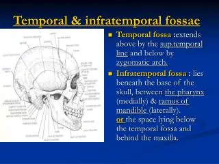
TEMPORAL & INFRATEMPORAL FOSSA I. Dr. Ahmed Fathalla Ibrahim. TEMPORAL FOSSA. TEMPORAL FOSSA. INFRATEMPORAL FOSSA. TEMPORAL FOSSA. BOUNDARIES: Anterior: Zygomatic process of frontal bone + zygomatic bone Superior & Posterior: Temporal lines Inferior: Zygomatic arch.

E N D
TEMPORAL & INFRATEMPORAL FOSSA I Dr. Ahmed Fathalla Ibrahim
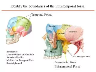
TEMPORAL FOSSA BOUNDARIES: • Anterior:Zygomatic process of frontal bone + zygomatic bone • Superior & Posterior:Temporal lines • Inferior:Zygomatic arch
TEMPORAL FOSSA CONTENTS: • Temporalis muscle • Deep temporal nerve & vessels • Superficial temporal vessels • Auriculotemporal nerve
INFRATEMPORAL FOSSA BOUNDARIES: • Superficial (lateral):Ramus of mandible • Deep (medial):Lateral pterygoid plate • Superior:Infratemporal surface of greater wing of sphenoid • Anterior:Tuberosity of maxilla
INFRATEMPORAL FOSSA COMMUNICATIONS: • With temporal fossa: through a gap deep to zygomatic arch • With cranial cavity: through foramen ovale, foramen spinosum, foramen lacerum • With orbit: through inferior orbital fissure • With pterygopalatine fossa: through pterygomaxillary fissure
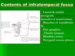
INFRATEMPORAL FOSSA CONTENTS: • Lateral & medial pterygoid muscles • Mandibular nerve & its branches • A part of maxillary nerve & 2 of its branches • Otic ganglion • Chorda tympani • A part of maxillary artery & 2 of its branches • Pterygoid plexus of veins
TEMPOROMANDIBULAR JOINT • TYPE: synovial, condylar variety • ARTICULAR SURFACES: • Upper: mandibular fossa + articular tubercle • Lower: head of mandible • CAPSULE: attached to: • margins of mandibular fossa & articular tubercle • Neck of mandible
TEMPOROMANDIBULAR JOINT • JOINT CAVITY: divided into upper & lower parts by an articular disc • ARTICULAR DISC: a plate of fibrocartilage with a concavoconvex upper surface and a concave lower surface • LIGAMENTS • Lateral temporomandibular: from tubercle on the root of zygoma to lateral surface of neck of mandible • Sphenomandibular: from spine of sphenoid to lingula of mandible • Stylomandibular
TEMPOROMANDIBULAR JOINT • NERVE SUPPLY: auriculotemporal • ARTERIAL SUPPLY: maxillary • MOVEMENTS: elevation, depression, protrusion, retraction, chewing (side-to- side) movements • RELATIONS • Anterior: mandibular notch, masseteric nerve & vessels • Posterior: tympanic plate of temporal bone • Lateral: Parotid gland • Medial: auriculotemporal nerve, maxillary artery
MUSCLES OF MASTICATION • TEMPORALIS • MASSETER • LATERAL PTERYGOID • MEDIAL PTERYGOID • They are derived from mesoderm of 1stbranchial arch • They originate from temporal or infratemporalfossa • They are inserted intoramus of mandible • They are supplied, through their deep surfaces by branches of mandibular nerve • They act ontemporomandibular joint
TEMPORALIS • ORIGIN: floor of temporal fossa & deep surface of temporal fasica • INSERTION: the tendon passes deep to zygomatic arch to be inserted to all coronoid process (except its lateral surface) • NERVE SUPPLY: 2 deep temporal nerves fromanterior division of mandibular nerve • ACTION: • Elevation of mandible • Its posterior fibers retract the mandible
MASSETER • ORIGIN: lower border & inner surface of zygomatic arch • INSERTION: lateral surface of ramus of mandible • NERVE SUPPLY: fromanterior division of mandibular nerve • ACTION: • Elevation of mandible • Protrusion of mandible (when muscles on both sides act together) • Side-to-side movement (when muscles on both sides act alternatively)
LATERAL PTERYGOID • ORIGIN: • Upper head: infratemporal surface of greater wing of sphenoid • Lower head: lateral surface of lateral pterygoid plate • INSERTION: pterygoid fovea (in front of neck of mandible) + capsule & articular disc of TMJ • NERVE SUPPLY: fromanterior division of mandibular nerve • ACTION: • Pulls the condylar process forward to depress the mandible • Protrusion of mandible (when muscles on both sides act together) • Side-to-side movement (when muscles on both sides act alternatively)
MEDIAL PTERYGOID • ORIGIN: • Superficial head: tuberosity of maxilla • Deep head: medial surface of lateral pterygoid plate • INSERTION: medial surface of ramus & angle of mandible • NERVE SUPPLY: fromtrunk of mandibular nerve • ACTION: • Elevation of mandible • Protrusion of mandible (when muscles on both sides act together) • Side-to-side movement (when muscles on both sides act alternatively)
ALL MUSCLES OF MASTICATION Elevate mandible EXCEPT • Lateral pterygoid Protrude mandible EXCEPT • Temporalis Are supplied by anterior division of mandibular nerve EXCEPT • Medial pterygoid
MANDIBULAR NERVE • COMPOSITION: • Formed of 2 roots: motor & sensory • ORIGIN: • Sensory root: peripheral processes of cells of trigeminal ganglion in the middle cranial fossa • Motor root: axons of cells of motor nucleus of trigeminal nerve in pons
MANDIBULAR NERVE • COURSE: • Both roots emerge separately through foramen ovale to infratemporal fossa • The 2 roots unite, below foramen ovale • The nerve soon divides into a small anterior & a large posterior division
MANDIBULAR NERVE • RELATIONS: • Superficial: lateral pterygoid • Deep: otic ganglion • Posterior: middle meningeal artery
BRANCHES FROM TRUNKOF MANDIBULAR NERVE • One motor:Nerve to medial pterygoid: supplies medial pterygoid & gives off 2 branches that pass through otic ganglion (without relay) & supply tensor palati & tensor tympani muscles • One sensory: meningeal branch (nervus spinosus) passing through foramen spinosum to supply meninges of middle cranial fossa
BRANCHES FROM ANTERIOR DIVISION OF MANDIBULAR NERVE Four branches Three motor: • Masseteric nerve: emerges through upper border of lateral pterygoid & turns along mandibular notch to reach masseter • Deep temporal nerves: emerge through upper border of lateral pterygoid • Nerve to lateral pterygoid One sensory: • Buccal nerve: emergesbetween the 2 headsof lateral pterygoid, supplies skin & mucous membrane overlying buccinator
BRANCHES FROM POSTERIOR DIVISION OF MANDIBULAR NERVE Four branches Three sensory: Auriculotemporal nerve: • Arises by 2 roots encircling middle meningeal artery • Runs backward, deep to neck of mandible • Gives sensory branches to skin of auricle, temple, TMJ & parotid gland • Carries postganglionic parasympathetic secretomotor fibers from otic ganglion to parotid gland
BRANCHES FROM POSTERIOR DIVISION OF MANDIBULAR NERVE Lingual nerve: • Emerges through lower border of lateral pterygoid then superficial to medial pterygoid • Joins chorda tympani • Runs just below 3rd molar tooth (dangerous position because it is only covered by muscous membrane) • Runs superficial to hyoglossus & is connected to submandibular ganglion by 2 roots • Carries general sensations from anterior 2/3 of tongue
BRANCHES FROM POSTERIOR DIVISION OF MANDIBULAR NERVE Inferior alveolar nerve: • Emerges through lower border of lateral pterygoid then superficial to medial pterygoid, behind lingual nerve • Passes through mandibular foramen & canal to supply lower teeth • Emerges through mental foramen as mental nerve supplying skin of lower lip & chin
BRANCHES FROM POSTERIOR DIVISION OF MANDIBULAR NERVE One motor: Mylohyoid nerve: • A branch of inferior alveolar nerve just above mandibular foramen • Passes in mylohyoid groove of mandible • Supplies mylohyoid & anterior belly of digastric muscles
RELATIONS OF LATERAL PTERYGOID • Superficial: temporalis, masseter, ramus of mandible, maxillary artery, buccal nerve • Deep: medial pterygoid, mandibular nerve, middle meningeal artery, otic ganglion • Emerging through its upper border: deep temporal & masseteric nerves • Emerging through its lower border: lingual & inferior alveolar nerves + maxillary artery • Emerging between its 2 heads: buccal nerve, maxillary artery