Download
1 / 25
320 likes | 646 Vues
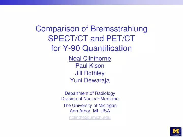
Comparison of Bremsstrahlung SPECT/CT and PET/CT for Y-90 Quantification. Neal Clinthorne Paul Kison Jill Rothley Yuni Dewaraja Department of Radiology Division of Nuclear Medicine The University of Michigan Ann Arbor, MI USA nclintho@umich.edu. Y-90 Imaging.

E N D
Comparison of Bremsstrahlung SPECT/CT and PET/CT for Y-90 Quantification Neal ClinthornePaul KisonJill RothleyYuni Dewaraja Department of RadiologyDivision of Nuclear Medicine The University of MichiganAnn Arbor, MI USA nclintho@umich.edu
Y-90 Imaging • Desirable for verifying distribution of Y-90 microspheres used in SIRT for unresectable liver cancer • Can potentially be used for dosimetry • Possible with both SPECT and PET • Goal was to compare performance of PET and SPECT using existing commercial acquisition and reconstruction
Phantom with Spheres Phantom having spheres with volumes ranging from 1.9ml (~1.5cm diameter) to 100ml (~5.7cm diameter) used for quantification studies 10000 ml 11.0 ml 63.0 ml 100.0 ml 1.9 ml
Initial Loading without Background • Spheres loaded with total of 341 MBq(9.2 mCi) of Y-90 chloride + EDTA • Y-90 assayed on Capintec CRC 15R using settings 55 (x10) for plastic syringe and 48 (x10) for glass vials • Scanned with PET and then SPECT 0 MBq 24.7 MBq 2.2 MBq/ml 199.0 MBq 2.0 MBq/ml 106.6 MBq 1.7 MBq/ml 5.3 MBq 2.8 MBq/ml
Spheres with Background 414.6 MBq 0.04 MBq/ml • One week (2.6 half-lives) later, 414.6 MBq (11.2 mCi) of Y-90 chloride was loaded into water surrounding spheres as background • Phantom rescanned with SPECT and then PET 4.1 MBq 0.37 MBq/ml 33.3 MBq 0.33 MBq/ml 17.8 MBq 0.28 MBq/ml 0.9 MBq 0.46 MBq/ml
SPECT Imaging Y-90 Energy Spectrum • Siemens Symbia T6 TruePoint SPECT/CT • High-energy collimators • Six contiguous energy windows 105–285 keV • 128x128 matrix 4.8mm pixels, OSEM (Flash 3D) 6 subsets, 20 iterations • No scatter correction • Attenuation correction from CT using center energy for each window Energy (keV)
PET Imaging • Siemens Biograph 6 extended FOV (21.6cm) LSO-based PET/CT • 435–650 keV window • Phantom imaged in one bed position • 3D OSEM reconstruction, 3 iterations, 21 subsets • No TOF • No copper or lead filter to moderate bremsstrahlung count rate • CT used for attenuation correction
Analysis • Spherical VOIs for spheres and background regions defined on CT scan for each dataset • For PET quantification, images were reconstructed to obtain radionuclide concentration in Bq/ml with F-18 calibration and corrected for Y-90 positron fraction • For SPECT, the largest (100 ml) sphere was used for calibration in each scan
SPECT 135–165 keV Window MIP 70 minute acquisition time
SPECT 255–285 keV Window MIP 70 minute acquisition time
PET No Background MIP 40 minute acquisition time (equivalent to SPECT with decay)
SPECT 135–165 keV Window + Bkgd MIP 120 minute acquisition time
SPECT 255–285 keV Window + Bkgd MIP 120 minute acquisition time
PET + Background MIP 120 minute acquisition time
SPECT Improvements • Shown in previous talk
PET Improvements • Time-of-flight • Shielding of low-energy bremsstrahlung? • More appropriate reconstruction • Better random coincidence modeling • Methods for reducing bias at low activities for datasets with few counts
Summary • Both PET and bremsstrahlung SPECT are usable for Y-90 imaging • SPECT with high-energy collimators has moderate resolution and suffers from partial volume effects for small sources but much higher countrate than PET • PET suffers from a low event rate, high background from bremsstrahlung, and significant positive bias in regions of low activity but has inherently higher resolution than SPECT • Both will likely be useful for quantitative Y-90 imaging if appropriate corrections are performed