Download
1 / 18
180 likes | 420 Vues
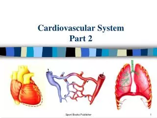
Cardiovascular System Part 2. Circulation Heart Anatomy Contraction Blood Pressure Cardiac Action Potentials EKG. Circulation. Pulmonary Circuit: R. atrium R. ventricle Pulmonary arteries Lung capillaries Pulmonary veins Systemic Circuit: L. atrium L. ventricle Aorta

E N D
Cardiovascular System Part 2 Circulation Heart Anatomy Contraction Blood Pressure Cardiac Action Potentials EKG
Circulation • Pulmonary Circuit: • R. atrium • R. ventricle • Pulmonary arteries • Lung capillaries • Pulmonary veins • Systemic Circuit: • L. atrium • L. ventricle • Aorta • Systemic arteries • Organ & tissue capillaries • Systemic veins • Vena cavas
Circulation • Atherosclerosis= cholesterol and lipid buildup on and in the walls of vessels. • Increased thickening increased pressure thickening decreased elasticity potential clotting
Circulation • Aneurysm= swelling or dilation of aorta or other blood vessels • May lead to rupture • May be caused by: • Aging breakdown of elastic fibers • Atheroscleosis • Genetic disorders • Infection
Heart Anatomy • Myocardium= cardiac muscle tissue • Pericardium= tough fibrous sac around the heart
Contraction • Heartbeat= one cardiac cycle; one contraction/ relaxation • Systole= contraction • Diastole= relaxation • Contraction of a chamber causes an increase in pressure that opens the valve to the next chamber. • “Lub-Dub” sound produced by the closing of AV and semilunar valves respectively.
Blood Pressure • Blood pressure= pressure exerted by the blood against the vessel walls • Normal = 120/80 • 120: systolic pressure; highest pressure • 80: diastolic pressure; lowest pressure • Hypertension= high blood pressure increased resistance to flow enlargement and weakening of heart
Cardiac Action Potentials • Self-excitatory pacemaker cells= • Spontaneously produce action potentials • Intercalated discs= • Bridge plasma membranes between 2 cardiac cells • Offer almost a spontaneous contraction of many cardiac cells at once
Cardiac Action Potentials • Pacemakers: • Sinoatrial (SA) node: • initiates atrial contraction • Sends signal from location in the R. atrium through the left atrium • Atrioventricular (AV) node: • Located in the septum • When SA signal is received at AV node there is a slow response • The AV node sends the signal down the purkinje fibers • Ventricles contract
EKG (aka ECG) • Electrocardiogram • Evaluates electrical events in the heart • Reads a potential difference between electrodes on the skin • Measures change in ECF currents, not in individual cells
EKG (aka ECG) • P-wave= atrial depolarization • QRS-complex= ventricular depolarization (w/atrial repolarization) • T-wave= ventricular repolarization
EKG (aka ECG) • Little to no summation in cardiac muscle: • No tetanic contraction • Refractory period: • Time after excitation when cells can not reach action potential • Allows for ventricular filling • Caused by the slow inactivation of sodium channels
EKG (aka ECG) • Arrhythmia= • Irregular heart rhythms • Pacemakers irregular • Detectable by EKGs • Ex) • Bradychardia – fewer beats/min than average • Tachychardia – many more beats/min than average • Ventricular fibrillation – haphazard contractions
EKG (aka ECG) More arrythmias: http://www.rnceus.com/course_frame.asp?exam_id=16&directory=ekg