Download
1 / 2
20 likes | 158 Vues
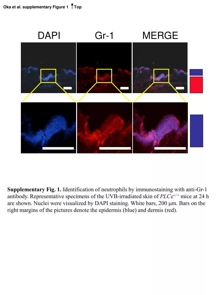
Oka et al. supplementary Figure 1 Top. DAPI. Gr-1. MERGE. Supplementary Fig. 1. Identification of neutrophils by immunostaining with anti-Gr-1 antibody. Representative specimens of the UVB-irradiated skin of PLC +/+ mice at 24 h

E N D
Oka et al. supplementary Figure 1 Top DAPI Gr-1 MERGE Supplementary Fig. 1. Identification of neutrophils by immunostaining with anti-Gr-1 antibody. Representative specimens of the UVB-irradiated skin of PLC+/+ mice at 24 h are shown. Nuclei were visualized by DAPI staining. White bars, 200 mm. Bars on the right margins of the pictures denote the epidermis (blue) and dermis (red).
Oka et al. supplementary Figure 2 Top +/+ -/- Supplementary Fig. 2. Effect of UVB irradiation on the SA-b-gal activity in the skin. PLC+/+ and PLC-/- mice (n = 4 for each genotype) were irradiated with a single dose of 1 kJ/m2 UVB. Frozen skin specimens were prepared 24 h after the irradiation, and SA-b-gal activity was determined by staining SA-b-gal using a senescence detection kit (BioVision Research Products, Mountain View, CA) accordingto the manufacturer’s instructions. Representative photographs of the skin from PLC+/+ (+/+) and PLC-/- (-/-) mice are shown. Bar, 100 mm. Dotted lines denote epidermis.