Download
1 / 26
340 likes | 934 Vues
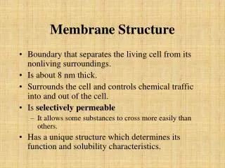
Membranes Structure of Membrane Proteins. The Manifold Roles of Membranes. The border of the cell/organelle Barrier to toxic molecules Helps accumulate & retain nutrients Carries out energy transduction Modulate signal transduction Mediate cell-cell interactions export of proteins

E N D
Membranes Structure of Membrane Proteins
The Manifold Roles of Membranes • The border of the cell/organelle • Barrier to toxic molecules • Helps accumulate & retain nutrients • Carries out energy transduction • Modulate signal transduction • Mediate cell-cell interactions • export of proteins • Facilitates cell motion • Assists in reproduction
Polar Head Group Hydrophobic Tail air water Spontaneously formed lipid structures • Like snowflakes, lipids usually travel in packs • Hydrophobic interactions are the key Monolayers arrange their hydrophobic tails in the air
Spontaneously formed lipid structures con’t Micelles bury the nonpolar tails in the center of a spherical structure nonpolar solvent Micelles reverse in nonpolar solvents water
Lipid bilayers: The basis for biological membranes multilamellar vesicles Still more spontaneously formed lipid structures unilamellar vesicles (liposomes)
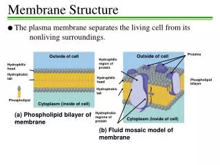
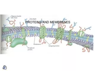
The Fluid Mosaic Model of Singer & Nicholson • The phospholipid bilayer is a fluid matrix • The bilayer is a two-dimensional solvent • Lipids and proteins can undergo rotational and lateral movement • Two classes of proteins: • peripheral proteins (extrinsic proteins) • integral proteins (intrinsic proteins)
Motion in the bilayer • Lipid chains can bend, tilt and rotate • Lipids and proteins can migrate ("diffuse") in the bilayer • Frye and Edidin proved this (for proteins), using fluorescent-labelled antibodies against membrane proteins (figure) • Lipid diffusion has been demonstrated as well
Membranes are Asymmetric • Lateral Asymmetry of Proteins: • Proteins can associate and cluster in the plane of the membrane - they are not uniformly distributed in many cases eg neurons • Lateral Asymmetry of Lipids: • Lipids can cluster in the plane of the membrane - they are not uniformly distributed • certain types may cluster around particular proteins in the membrane (a lipid entourage) • Can also induce asymmetry eg with calcium ion treatment
Transverse Asymmetry of Membranes • Functions of membrane proteins depends on their orientation within the membrane • Membrane proteins are not tossed into the membrane randomly but have a specific topology • eg Glycophorin Outside Inside
Transverse asymmetry of membrane lipids In most cell membranes, the lipid composition of the outer monolayer differs from that of the inner monolayer: phosphatidylcholine phosphatidyethanolamine phosphatidylserine sphingomyelin total percentage of phospholipid
How to establish & maintain lipid asymmetry?The role of flippases • Lipids can be moved from one monolayer to the other by flippase proteins • Some flippases operate passively and do not require an energy source • Other flippases appear to operate actively and require the energy of hydrolysis of ATP
Membrane Phase Transitions: The "melting" of membrane lipids • Below a certain transition temperature, membrane lipids are rigid & tightly packed (Gel Phase) • Above the transition temperature, lipids are more flexible and mobile (Liquid crystal phase) • The transition temperature is characteristic of the lipids in the membrane • Only pure lipid systems give sharp, well-defined transition temperatures Liquid crystal Gel
Transition Temperature Liquid crystal Anti conformation Gel Heat absorbed Gauche conformations Temperature Observing Membrane Phase Transitions by Calorimetry
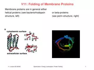
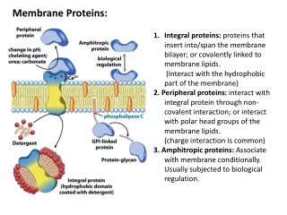
Integral (intrinsic) proteins • Peripheral (extrinsic) proteins • Lipid-anchored proteins Structure of Membrane Proteins
Peripheral Membrane Proteins • Not strongly bound to the membrane • Can be dissociated with mild detergent treatment or with high salt concentrations • What holds them there in the first place?
Integral Membrane Proteins (IMPs) • Strongly imbedded in the bilayer • Can only be removed by disrupting the membrane (eg detergents) • Often span the membrane (transmembrane) • “Inside out” compared to other globular proteins..meaning?
Roles of IMPs (will expand on next term & beyond) • Identification of cell type (‘face’) • Structural eg adhesion proteins • Signalling: eg mediate cell growth & differentiation • Pumps & Channels: import & export control
Bacteriorhodopsin: a classic example of a serpentine IMP • Function: a light-driven proton pump • Consists of 7 transmembrane helical segments with short loops that interconnent the helices • Binds a light-senstive cofactor (retinal) in the hydrophobic core • Found in purple patches of Halobacterium halobium
Porins: Classic example of a “b-basket” IMP • Function: act as selective pores for various small molecules • Structure: ~ membrane-spanning 18-strand b barrel forming a hollow cylinder • Interior of cylinder lined with hydrophilic residues • Found in outer membrane of Gm- bacteria & mitochondria eg maltoporin
Lipid-Anchored Proteins • Relatively new class of membrane proteins • Four types have been found: • Amide-linked myristoyl anchors • Thioester-linked fatty acyl anchors • Thioether-linked prenyl anchors • Glycosyl phosphatidylinositol anchors In Out Critical for proper protein function: location, location, location! Often found on proteins involved in signal transduction
Amide-Linked Myristoyl Anchors • Anchor is myristic acid (what is the abbreviated name?) • Myristic acid forms an amide linkage with the protein at its amino terminus • N-terminal residue is always glycine • Examples: • a subunits of G proteins endothelial nitric oxide synthase signal transduction
Ester-linked Acyl Anchors • Broad specificity for lipids - myristate, palmitate, stearate, oleate all found • Broad specificity for amino acid links - Cys, Ser, Thr all found • e.g G-protein-coupled receptors, Transferrin receptor
! N-myristoylation S-palmitoylation
Thioether-linked Prenyl Anchors • Prenylation refers to linking of "isoprene"-based groups • Consensus sequence CAAX (C=Cys, A=Aliphatic, X= any) • Isoprene groups include farnesyl (15-C) & geranylgeranyl (20 C) groups • e.g yeast mating factors, intracellular signalling proteins
eg farnesylation What’s the Point? • Lipid Anchors are Signaling Devices • Anchors are transient • Reversible anchoring and de-anchoring can control signalling pathways • (Similar to phosphorylation/ dephosphorylation, substrate binding/ dissociation, proteolytic cleavage triggers and signals)
Protein Glycosyl Phosphatidylinositol Anchors (GPI anchors) O CO C • Anchors protein lying outside the cell • Always attached to aC-terminalresidue • Ethanolamine linked to a phosphate linked to an oligosaccharide linked in turn to inositol of phosphatidyl inositol (embedded in the membrane) • Examples: surface antigens, adhesion molecules, cell surface hydrolases HNCH2CH2OPO- O (Man)3 GN PI Man = mannose GN = glucosamine PI = phosphatidyl inositol