Download
1 / 23
250 likes | 759 Vues
Chemical synapses: post-synaptic mechanisms. Postsynaptic Membranes and ion channels . Ligand gated ion channels – a review. Resting K + channels : responsible for generating the resting potential across the membrane

E N D
Postsynaptic Membranes and ion channels • Ligand gated ion channels – a review • Resting K+ channels: responsible for generating the resting potential across the membrane • Voltage- gated channels: responsible for propagating action potentials along the axonal membrane • Two types of ion channels in dendrites and cell bodies are responsible for generating electric signals in postsynaptic cells. • (c) Has a site for binding a specific extracellular neurotransmitter • (d) Coupled to a neurotransmitter receptor via a G protein.
More things to know about Ion channels • All the ion channels in question have a common feature • A pore that allows the ion(s) in question to flow across the lipid bilayer • The pore is specific to a certain ion or ions • Leak K+ channel only allows K+ ions to flow across the membrane • Example: Acetylcholine (ACH) receptor allows Na+ to flow and the glycine receptor allows Cl- to flow through the channel • Ligand gated ion channels are different than voltage-gated ion channels in that they are chemically gated ie via neurotransmitters • Binding a small chemical triggers the opening of the ion channel • i) Na+ channels - excitatory (generates an excitatory postsynaptic potential)ii) Cl- channels - inhibitory (generates an inhibitory postsynaptic potential) • Important: The specificity of a transmitter response is a function of the receptor type NOT the transmitter itself. (i.e. Ach can be excitatory when binding to one type of AchR (NMJ)) and inhibitory when binding to another type of receptor
Acetylcholine – general info • Motor neuron transmitter at the neuromusccular junction (NMJ) in vertebrates • Present in brain (10% of synapses) • Packaged in high numbers in vesicles 1,000 to 10,000 molecules per vesicle at the NMJ • Like all small chemical transmitters Ach is synthesized and packaged into vesicles in the synapse • The NMJ pre-synaptic side is packed full of vesicles in the axon terminal • Many vesicles are released per action potential to ensure a large safety margin so that the muscle fiber (i.e. the postsynaptic cell) will depolarize to beyond threshold.
Acetylcholine – receptor • Officially called the nicotinic ACH receptor (nAChR) because nicotine binds to this receptor and activates it • ligand gated ion channel • has a depolarizing effect because Na+ is the dominant ion through these channels
Acetylcholine – receptor • generates an excitatory postsynaptic potential which at the NMJ (motor end plate) is often called an "end plate potential“
EPP - end plate potential • Aka Excitatory Junctional Potential (EJP) End plate potentials (EPPs) evoked by stimulation of a motor neuron are normally above threshold and therefore produce an action potential in the postsynaptic muscle cell.
nACHR – a closer look • Most of the mass of the protein protrudes from the outer (synaptic) surface of the plasma membrane • The M2 alpha helix (red) in each subunit is part of the lining of the ion channel • Aspartate and glutamate side chains at both ends of each M2 helix form two rings of negative charges that help exclude anions from and attract cations to the channel. The gate, which is opened by binding of acetylcholine, lies within the pore. Aspartate
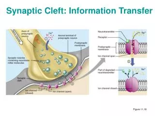
The Neuromuscular junction • Arrival of an action potential at the terminus of a presynaptic motor neuron induces opening of voltage-gated Ca2+ channels • subsequent release of acetylcholine, which triggers opening of the ligand-gated nicotinic receptors in the muscle plasma membrane • The resulting influx of Na+ produces a localized depolarization of the membrane • leading to opening of voltage-gated Na+ channels and generation of an action potential * * *
Synapses in the brain or central nervous system (CNS) • A single synapse on a target is seldom found in brain • Large neurons in the brain typically receive many inputs (1000 to 80,000 per cell) • The inputs are integrated in the receiving neuron such that a "decision" is made to pass on the information onto other cells - this "decision" is often whether or not to generate an action potential • each synaptic input usually only gives only a small depolarization, so many inputs must cooperate (summate) to reach threshold to fire an action potential
EPSP • An excitatory impulse, an excitatory post-synaptic potential raises the membrane potential above rest • 1. An excitatory impulse at a synapse on the soma causes a depolarization of the whole soma including the beginning of the axon. This is because the diameter of the soma or cell body is so large that the internal resistance is very low so current flow extremely well through the cell body • The beginning of the axon is also known as the spike initialization zone or axon hillock and is packed with Na+ channels, an epsp of +15 to +20 mV triggers an action potential in the zone • 2. An epsp generated on a dendrite will diminish in strength by the time the current has reached the soma such that an epsp in a dendrite has less of a chance of triggering an action potential than an epsp generated at the soma • Due to the absence of voltage-gated Na+ channels in the soma and dendrite of most neurons it is very unlikely that an action potential will be generated in these regions
IPSP • An inhibitory impulse is called an ipsp (inhibitory post-synaptic potential) and lowers the membrane potential below rest (hyperpolarizes) • Synaptic transmission triggers the opening ligand gated Cl- channels or indirectly through other mechanisms the opening of K+ channels • Cl- flows into the cell • K+ flows out of the cell • Both increase the negative charge within the cell, hyperpolarizes the soma • Brings membrane potential further away from threshold and so it is harder to trigger an action potential therefore inhibitory • An ipsp on the dendrite will have less effect due to current loss than an ipsp in the soma
CNS • Major ligand gated ion channels and their neurotransmitters • Glutamate - amino acid • Most common excitatory neurotransmitters in central nervous system • Neurotransmitter of NMJ in invertebrates (locust, giant axon of squid) • Glutamate receptor - at least 3 different ligand gated ion channel receptors for glutamate - all generate epsps as Na+ is the dominant ion that flows after the channel is open • GABA - aminobutyric acid • Major inhibitory neurotransmitter in the brain • In some areas of cortex 1 in 5 neurons are GABAergic • GABA receptors- again many different types of receptors - the more common GABA receptors are Cl - channels- usually inhibitory causes an inhibitory postsynaptic potential (IPSP)- reversal potential is the same as ECl - usually around - 70 mV • Note: reversal potential is synonymous with equilibrium potential • cont…..
CNS • Glycine - simplest amino acid • Major inhibitory neurotransmitter in the brainstem and spinal cord • Glycine Receptor- major receptor is a Cl - channel- inhibitory- like GABA receptor in that usually causes IPSPs- blocked by strychnine (rat poison) which literally causes convulsions and death as now the motor neurons are not inhibited and the muscles contract without control. Yikes!
Cable properties again • Dendrites extend 0.5 to 1 mm in all directions from soma and receive signals from a large area • 80-90% of all presynaptic terminals terminate on dendrites • Most can't produce action potentials (too few or no Na+ channels) • Transmit current by passive spread down dendrites to the soma • Therefore the membrane potential decreases as move along dendrite due to current loss thanks to our friends ri, rm and cm • Dendrites have no voltage gated Na+channels and cell bodies ie soma have little or no voltage-gated Na+ channels current flow is solely dependent on the Cable Properties of the dendrites and soma
Things to remember • 1) loss of current across membrane (leaky membranes) • dependent on the internal resistance (ri) and the membrane resistance (rm) • the length or space constant describes this property • 2) loss of current (charge) due to capacitance properties of the membrane • cell membrane acts as a capacitor • it takes time and current (charge) to charge the membrane capacitor • the time constant describes this effectτ = Rm x Cm • details are in the lecture on cable properties
Summation - CNS • The postsynaptic effects of most synapses in the brain are not as large as those at the neuromuscular junction • In the CNS the postsynaptic potentials are usually far below the threshold for generating postsynaptic action potentials • Neurons in the central nervous system are typically innervated by thousands of synapses, and the postsynaptic potentials produced by each active synapse can summate together (in space and in time) to bring the membrane to threshold for firing an action potential
Motor neurons and summation • Each motor neuron synapses with multiple muscle fibers • The motor neuron and the fibers it contacts defines the motor unit
Summation Summation of multiple epsps to bring the membrane potential to threshold for an action potential.
Summation • A microelectrode records the postsynaptic potentials produced by the activity of two excitatory synapses (E1 and E2) and an inhibitory synapse (I) • Electrical responses to synaptic activation • Stimulating either excitatory synapse (E1 or E2) produces a subthreshold EPSP, whereas stimulating both synapses at the same time (E1 + E2) produces a suprathreshold EPSP that evokes a postsynaptic action potential • Activation of the inhibitory synapse alone (I) results in a hyperpolarizing IPSP • Summing this IPSP with the EPSP produced by • one excitatory synapse (E1 + I) reduces the • amplitude of the EPSP, while summing it with the • suprathreshold EPSP produced by activating • synapses E1 and E2 keeps the postsynaptic • neuron below threshold, so that no action • potential is evoked.