Download
1 / 14
140 likes | 254 Vues
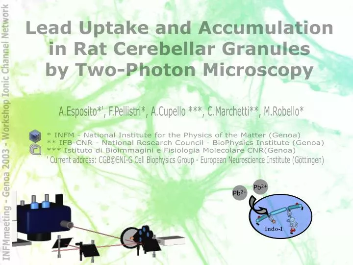
Pb 2+. Pb 2+. Lead Uptake and Accumulation in Rat Cerebellar Granules by Two-Photon Microscopy. Title. A.Esposito*', F.Pellistri*, A.Cupello ***, C.Marchetti**, M.Robello*. * INFM - National Institute for the Physics of the Matter (Genoa)

E N D
Pb2+ Pb2+ Lead Uptake and Accumulation in Rat Cerebellar Granules by Two-Photon Microscopy Title A.Esposito*', F.Pellistri*, A.Cupello ***, C.Marchetti**, M.Robello* * INFM - National Institute for the Physics of the Matter (Genoa) ** IFB-CNR - National Research Council - BioPhysics Institute (Genoa) *** Istituto di Bioimmagini e Fisiologia Molecolare CNR(Genoa) ' Current address: CGB@ENI-G Cell Biophysics Group - European Neuroscience Institute (Göttingen)
Lead introduction Ca2+ Calcium, a second messenger Pb2+ Lead, interferes with calcium signalling Ca2+ Pb2+ Uptake machinery Lead action Pb2+ Pb2+ Ca2+ ? Lead Methods Results Discussion Lead H.A.Godwin, Curr.Opinion Chem.Bio. 5, 223-227 (2001) M.Mazzolini,S.Traverso,C.Marchetti, J.Neurochem.2001 Oct;79(2):407-16 2
Pb2+ Pb2+ Pb2+ Lead Methods Results Discussion Methods Probe, model and microscopy choice Ratcerebellar granule cells Membrane permeant high affinity fluorescent dye: Indo-1 Two-photon laser scanning microscopy (TPLSM) 45´ Indo-1 AM - 1M 30´ Standard solution @37°C 3 TPEN (100M)
Pb2+ Lead Methods Results Discussion Methods Indo spectra and binding Autofluorescence <<1% Residual Indo1 fluorescence: <5% M.E.Legare, R.Barhoumi, E.Hebert, G.R.Bratton, R.C.Burghardt and E.Tiffany-Castiglioni, “Analysis of Pb2+ Entry into Cultured Astroglia”, Toxicological Sciences 46, 90-100 (1998) 4 Figura ‑5 a) rappresentazione dell’eccitazione a doppio fotone di una molecola di Indo-1. b) spettro dell’Indo-1 a varie concentrazioni di calcio, si nota una lunghezza d’onda non sensibile alla variazione di tale ione; c) sezioni d’urto d’azione dell’Indo-1. In azzurro la sonda libera, in violetto quella legata; d) Spettri relativi della sonda libera (in azzurro), legata al calcio (in violetto) e legata al piombo (in nero). Si evidenziano i picchi a 405nm (punto a) e 485nm (punto b) corrispondenti rispettivamente alla sonda legata al calcio e libera. Inoltre a 460nm (punto c) il punto isosbestico, insensibile alla concentrazione del calcio, ma dipendente da quella del piombo (d).
TPLSM Lead Methods Results Discussion Methods Indo-1 UV excitation Reduced photoxicity and photodamages High resolution and confocality TPM Resolution Axial: 700nm Lateral:350nm Temporal: 3s/pxl Wavelength: 700nm (680-1050nm) Mean power on the sample: 5-7mW Pulsed laser: 80MHz ; 100-200fs A.Diaspro, “Confocal and Two-Photon Microscopy”, Wiley-Liss (New York, 2002) F.M. Wahl, “Digital Image Signal Processing”, Artech House (1987) 5
Microscope Microscope TE300Eclipse (Nikon) PMT1 PMT2 Scanning Head (PCM2000) Tsunami (Spectra Physics) Millennia Lead Methods Results Discussion Methods A.Diaspro et al. (1999b), “Adapting a compact confocal […]”, Microsc. Res. Tech. 47, 196-205 A.Diaspro […] M.Robello and F.Olivini (2001), “Two-photon microscopy […]”, J.Biomed.Opt. 6
KCl 75mM La3+ (25M) 2‘ Fluorescence intensity Pb2+ (20M) TPEN (100M) time Lead Methods Results Discussion Methods Protocol 7
photobleaching quenching S1 T1 photochemistry S0 FLUORESCENCE time PHOSPHO_ RECENCE (3.40.5)10-4 % s-1 mW-2 frm-1 Bleaching rate ~3-8°/00frm-1 Fluorescence variations 2 control frames Photobleaching curve Bleaching estimation 2 4 6 0 8 -0% Time (min) -10% -20% Lead Methods Results Discussion Results Photobleaching 8
Indo1 concentration calibration 0 255 Intensity (a.u.) 5mm Lead Methods Results Discussion Results Autofluorescence <<1% Mean Indo-1: ~90mM 50/100mM Brust-Mascher and Webb, “Calcium Induced by Large Voltage Pulses in Fish Keratocytes”,Biophy.J. Vol75 1998 9
Var: 54±6% Raw: 67±9% Sat: 63±7% 75 Pb2+ (20µM) Pb2+ (20µM) 65 55 45 35 Estimated concentration (µM) 25 ~ 15 bound 5 0 2 4 Time (min) Lead Methods Results Discussion Results Lead uptake Time (min) 0 2 4 6 8 0% -15% -30% Fluorescence variations (%) -45% Pb2+(20µM) -60% Fluorescence variations Photobleaching -75% 10
75 Pb2+ (20µM) Pb2+ (20µM) 65 M) 55 Pb2+ 45 35 Concentration ( La3+ 25 15 +La3+ (25µM) 5 0 2 4 Time (min) Lead Methods Results Discussion Results Lanthanum inhibition 11
Pb2+ (20µM) Pb2+ (20µM) Lead Methods Results Discussion Results Free cytosolic lead 12
Conclusions Rapid lead uptake in the micromolar range (bound) Free cytosolic lead in the picomolar range Probable saturation of transport Partial lanthanum inhibition (~70%) Pb2+ Pb2+ Pb2+ Pb2+ Pb2+ La3+ C2 (PKC 2pM, sinaptogamin) EF-hand helic (calmodulin, calcineurin @ 100pM) Cys3(zinc proteins in pM range) Pb2+ Pb2+ Pb2+ Pb2+ Pb2+ Lead Methods Results Discussion Discussion R.H.S.Westerink et al., “Ca2+-indipendent vesicular catecholamine release in PC12 cells by nanomolar concentrations of Pb2+”, Journal of Neurochemistry, 2002 Vol.80 861-873 13
Aknoledgment Acknowledgment Acknowledgment Mauro Robello Carla Marchetti Francesca Pellistri Aroldo Cupello Alberto Diaspro Paola Ramoino Camilla Luccardini Silvia Casagrande Marzia Pisciotta Marco Raimondo Federico Federici Mauro Robello Carla Marchetti Fred Wouters Cell Biophysics Group European Neuroscience Institute Göttingen - Germany www.eni.gwdg.de Contact Contact Alessandro Esposito - PhD stud. @ ENI-G (CGB) FLIM (FD/TD) development, Molecular Biology www.gwdg.de/~aesposi aesposi@gwdg.de