Download
1 / 136
1.43k likes | 1.86k Vues
9. The Nervous System: Part One: The Information Super Highway. Multimedia Asset Directory. Slide 36 Multiple Sclerosis Animation Slide 46 Neurosynapses Video Slide 51 Muscle Contraction Animation Slide 64 Epidural Placement Video Slide 80 Spinal Cord Anatomy Animation

E N D
9 The Nervous System: Part One: The Information Super Highway
Multimedia Asset Directory Slide 36 Multiple Sclerosis Animation Slide 46 Neurosynapses Video Slide 51 Muscle Contraction Animation Slide 64 Epidural Placement Video Slide 80 Spinal Cord Anatomy Animation Slide 81 Brachial Plexus Animation Slide 82 Lumbrosacral Plexus Animation Slide 85 Cervical Spine Injuries Video Slide 91 Reflex Arc Animation Slide 113 Carpal Tunnel Syndrome Video Slide 114 Electroneurodiagnosticians Video
Introduction • The nervous system is complex and important to the body’s control system. • The nervous system monitors conditions and takes corrective action, when necessary, to keep everything running smoothly.
Introduction • The control systems of your body are the nervous and endocrine systems which receive help from your special senses. • Like any control system, they have a large, complex job that is sometimes difficult to understand. Thus, the systems themselves are perhaps the most complex and vital systems.
Learning Objectives • List and describe the components and basic operation of the nervous system. • Contrast the central and peripheral nervous systems. • Define the parts and functions of the nervous tissue. • Discuss the anatomy and physiology of the spinal cord. • List and describe various disorders of the nerves and spinal cord.
arachnoid mater (ah RAK noyd MAY ter) astrocytes (ASS troh SITES) axon (AK sahn) cerebrospinal fluid (SER eh broh SPY nal) chemical synapse (SIN naps) commissures (KAHM ih shoorz) corticobulbar tract (KOR ti coe BUL bar) corticospinal tract (KOR ti coe SPY nal) dendrites (DEN drights) Pronunciation Guide Click on the megaphone icon before each item to hear the pronunciation.
dorsal root ganglion (GANG lee on) dura mater (DOO rah MAY ter) ependymal cells (eh PEN deh mall) epidural space (EPP ih DOO rall) ganglia (GANG lee ah) glial cells (GLEE all) gyri (JIE rie) meninges (men IN jeez) microglia (my KROG lee ah) Pronunciation Guide Click on the megaphone icon before each item to hear the pronunciation.
myelin (MY eh lin) neuroglia (glial cells) (noo ROG lee ah) nodes of Ranvier (ron vee AYE) oligodendrocytes (AH li go DEN droe sites) pia mater (PEE ah MAY ter) plexus (PLECK sus) Schwann cells (SHWAN) somatic nervous system (so MAT ick) spinocerebellar tract (SPY no ser eh BELL ar) Pronunciation Guide Click on the megaphone icon before each item to hear the pronunciation.
spinothalamic tract (SPY no thol AH mic) subarachnoid space (SUB ah RACK noyd) subdural space (sub DOO ral) sulcus (SULL cus) vesicles (VESS ih kulz) Pronunciation Guide Click on the megaphone icon before each item to hear the pronunciation.
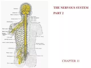
Parts and Basic Operations • The brain and spinal cord is the central nervous system (CNS) which controls the total nervous system. • Everything outside the brain and spinal cord is part of the peripheral nervous system (PNS). • The input side of the nervous system is the sensory system. • The output side of the nervous system is the motor system.
Parts and Basic Operations • The somatic nervous system controls skeletal muscle and mostly voluntary movements. • The autonomic nervous system controls smooth muscle and cardiac muscle, along with several glands. • The autonomic system is divided into the parasympathetic system that deals with normal body functioning while the sympathetic nervous system controls the “fight or flight” response system.
Real Life Example • You park your car and get out to visit a friend. As you step on the walk a large dog bounds down the steps barking and snarling at you. • Your sensory system gathers information including; a large unfriendly dog, you are far from the protection of your car, and no one is around to help. • The information goes into your spinal cord and brain and you process the information to make decisions. You are in danger; something must be done!
Real Life Example • Your CNS sends directions to your organs to gear up for action via the autonomic nervous system. • Your heart rate, blood pressure, and respiration rate increase. You begin to sweat. More blood is delivered to your skeletal muscles and heart in order to get you fully ready to respond. This is all involuntary, meaning you cannot consciously control it.
Real Life Example • Your somatic nervous system readies your skeletal muscles to get you out of there. This is often called the “fight or flight” response and will be discussed later in further depth. If you can control your fear, you back slowly away from the situation. If you are scared witless, you run from the yard as fast as possible. Either way, you can hopefully escape the danger, with your skin and pride intact.
Neuroglia • Specialized cells in the nervous system called neuroglia, or glial cells, perform specialized functions. • In the CNS there are four types of glial cells • Astrocytes – metabolic and structural support cells • Microglia – remove debris • Ependymal cells – cover and line cavities of the nervous system • Oligodendrocytes – make a lipid insulation called myelin
Neuroglia • In the PNS there are two types of glial cells: • Schwann cells – make myelin for the PNS • Satellite cells – support cells
Neurons • All of the control functions of the nervous system must be carried out by a group of cells called neurons. • Neurons have many branches and even what appears to be a tail.
Neurons • Each part of a neuron has a specific function • Body – cell metabolism • Dendrites – receive information from the environment • Axon – generates and sends signals to other cells • Axon terminal – where the signal leaves the cell • Synapse – where the axon terminal and receiving cell meet
Classification of Neurons • Neurons can be classified by how they look (structure) • Unipolar – 1 process with a peripheral and central projection • Bipolar – 2 processes, 1 axon and 1 dendrite • Multipolar – many processes • Or what they do (function) • Input neurons are known as sensory neurons. • Output neurons are known as motor neurons. • Neurons which carry information between neurons are called interneurons (inter – between) or association neurons.
How Neurons Work • Neurons are a kind of cell called an excitable cell. This simply means that if the cell is stimulated it can carry a small electrical charge. • Each time charged particles flow across a cell membrane, there is a tiny charge generated. • All three muscle types are excitable cells, as are many gland cells.
How Neurons Work • Cells are like miniature batteries, able to generate tiny currents simply by changing the permeability of their membranes.
Action Potential • A cell that is not stimulated or excited is called a resting cell; it is said to be polarized. • It has a difference in charge across the membrane, being more negative inside than outside the cell. • When the cell is stimulated: • Gates (called sodium gates) in the cell membrane spring open allowing sodium to travel across the membrane.
Action Potential • When the cell is stimulated: • These sodium bits are positively charged, so the cell becomes more positive as they enter. • A cell that is more positive is called depolarized. • The sodium gates close. • Potassium gates open and potassium leaves the cell, taking its positive charge with it. This is called repolarization.
Action Potential • When the cell is stimulated: • If the cell becomes more negative than resting it is called hyperpolarized. • Action potential (AP) is the cell moving through depolarization, repolarization, and hyperpolarization. • The cell cannot accept another stimulus until it returns to its resting state, and this time period when it cannot accept another stimulus is called the refractory period.
Local Potentials • Neurons can use their ability to generate electricity to send, receive, and interpret signals. • If you hit your thumb with a hammer, dendrites in your thumb are stimulated by the blow and sodium gates open, sodium flows into the dendrites and they become depolarized. The number of cells affected depends on how hard you hit your thumb.
Local Potentials • In local potential the size of the stimulus determines the excitement of the cell. Many sensory cells work via local potentials, which is how your CNS determines the size of the environmental change. • The dendrites carry the depolarization to the sensory neuron cell body, which takes the information and generates an action potential if the stimulus is big enough.
Local Potentials • One difference between action potentials and local potentials is that action potentials are “all-or-none,” meaning the depolarization always finishes and is always the same size, while local potentials vary in size depending on the stimulus.
Impulse Conduction • The speed of impulse conduction is determined by the amount of myelin and the diameter of the axon. • Myelin is a lipid insulation or sheath formed by the oligodendrocytes in the CNS and Schwann cells in the PNS. • Myelinated nerves look white while unmyelinated nerves are gray.
Impulse Conduction • Myelin is essential for speedy flow of AP’s down the axons. In an unmyelinated axon, the AP can only flow down the axon by depolarizing each and every centimeter of the axon (a relatively slow process). In myelinated axons there are nodes located periodically, and only the nodes must depolarize, allowing the impulse to travel quickly as it skips from node to node.
Clinical Application: Multiple Sclerosis • Multiple sclerosis (MS) is a disorder of the myelin in the CNS. Many areas of myelin are destroyed. In these areas, impulse conduction is slow or impossible. Symptoms of MS differ depending on where the myelin damage occurs. Disturbances in balance, vision, speech, or movement is possible. MS occurs more in women, and patients are usually under 50.
Click here to view a video on the topic of Multiple Sclerosis. Back to Directory
Impulse Conduction and Diameter • The diameter of the axon also affects the speed of the AP flow. The wider the diameter of the axon, the faster the flow of ions. • Myelination and larger diameters allow for a huge difference in speed.
Impulse Conduction and Diameter • Small unmyelinated axons have speeds as low as 0.5 meters/second while large-diameter myelinated axons may be as fast as 100 meters/second. That’s 200 times faster!!
How Synapses Work • When the AP arrives at the axon terminal, the terminal depolarizes and calcium gates open. Calcium flows into the cell. When calcium flows in, it triggers a change in the terminal.
How Synapses Work • There are tiny sacs in the terminal called vesicles which release their contents from the cell when calcium flows in. These vesicles are filled with molecules, called neurotransmitters, used to send the signal from the neuron across the synapse to the next cell in line.
Neurotransmitters • The neurotransmitters bind to the cell receiving the signal, opening or closing gates. Some excite the receiving cell and some calm it down. • The last step in the transfer of information is to clean up, removing the neurotransmitter from the synapse to prevent it from binding to the receiving cell.
Neurotransmitters • This type of synapse is called a chemical synapse because neurotransmitters carry the information from one cell to another.
Figure 9-6 The Chemical Synapse. Step 1: The impulse travels down the axon. Step 2: Vesicles are stimulated to release neurotransmitter (exocytosis). Step 3: The neurotransmitter travels across the synapse and binds with the receptor site of post synaptic cell. Step 4: The impulse continues down the dendrite.
Chemical Synapses andMedications • Our understanding of chemical synapses has lead to several breakthroughs for treating mental illness. • Many medications on the market today are designed to modify synapses.
Chemical Synapses andMedications • Selective serotonin reuptake inhibitors (SSRIs) are good examples. These medications prevent the clean up of the neurotransmitter serotonin from synapses, thus increasing the effects of serotonin on the receiving cell. • Many antidepressants and anti-anxiety drugs are SSRIs.
Click here to view a video on the topic of Synapses. Back to Directory
Electrical Synapses • Some cells do not need the chemicals to transmit information from one cell to another. • These synapses are electrical synapses, transferring information freely because they have special connections called gap junctions. • These kinds of connections can exist between any types of excitable cells. • They are found in the intercalated discs between cardiac muscle fibers.
The Neuromuscular Junction • The neuromuscular junction is a chemical synapse creating a specialized synapse between somatic (voluntary) motor neurons and the skeletal muscles they innervate. • The surface of the muscles is studded with sodium channels that are ligand gated. These open or close when a molecule binds to a receptor that is part of the channel, like a key fitting into a lock.
The Neuromuscular Junction • In the case of skeletal muscles, the ligand is the neurotransmitter acetylcholine, which is released from the terminal of a motor neuron and binds to the surface of skeletal muscle, opening sodium channels and causing the skeletal muscle to depolarize. The muscle then contracts. • Acetylcholinesterase is the enzyme responsible for cleaning up the synapse.