Download
1 / 30
390 likes | 1.19k Vues
Chapter 7 Cartilage. Cartilage is a specialized form of connective tissue. C ells: chondrocytes Extensive extracellular matrix. Structure:. ★ Chondrocytes synthesize and secrete extracellular matrix. ★ The chondrocytes are located in matrix cavities, called lacunae. Functions:.

E N D
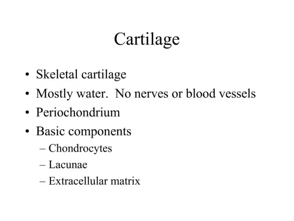
Cartilage is a specialized form of connective tissue.
C ells: chondrocytes Extensive extracellular matrix Structure: ★Chondrocytes synthesize and secrete extracellular matrix. ★The chondrocytes are located in matrix cavities, called lacunae.
Functions: ★to bear mechanical stresses without permanent disortion. ★to support soft tissue. ★to be a shocking-absorbing and sliding areas for joints and facilitate bone movements. ★to be essential for the development and growth of long bones both before and after birth.
More Features of Cartilage : ·Cartilage is avascular. ·Cartilage is nourished by the diffusion of nutrients from capillaries in adjacent connective tissue or by synovial fluid from join cavities. ·Chondrocytes exhibit low metabolic activity. ·Cartilage has no lymphatic vessels or nerves. ·Cartilage is surrounded by perichondrium in most places.
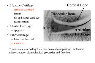
Classification: Hyaline cartilage: Elastic cartilage: Fibrocatilage: most common, much type II collagen elastic fibers, type II collagen dense network of type I collagen; high stress and weight bearing
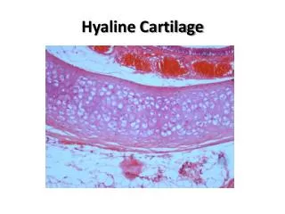
Hyaline cartilage ★Fresh hyaline cartilage is bluish-white and translucent. ★In the embryo, it serves as a temporary skeleton until it is gradually replaced by bone. ★In adult mammals, hyaline cartilage is located : in the articular surfaces of the movable joins; in the walls of large respiatory passage; in the ventral ends of ribs; in the epiphyseal plate.
Chondrocytes Matrix Perichondrium Strcture: Hyaline cartilage
①Collagen molecules ②Proteoghycans ③ Chondronectin Hyaline cartilage 1. Matrix (1) Hyaline cartilage matrix is produced by chondrocytes and contains three major classes of molecules:
①Collagen molecules Hyaline cartilage contains primarily type II collage. Small amounts of collagen types IX, X, XI, and others are frequently present.
②Proteoghycans chondroitin sulfate keratan sulfate hyaluronic acid forming proteoglycan aggregates.
③Chondronectin Chondronectin binds specifically to glycosaminoglycans and collagen type II, mediating the adherence of chondrocytes to the extracellular matrix.
(2) Hyaline cartilage matrix is highly hydrated to permit diffusion of small metabolites and resilience. Sixty and eighty percent of the net weight of hyaline cartilage is water. Water is loosely enough to allow diffusion of small metabolities to and from the chondrocytes. The high degree of hydration and the movement of water in the matrix allow the cartilage matrix to respond to varying pressure loads and contribute to cartilage’s weight-bearing capacity.
(3) Grounds substance components of hyaline cartilage matrix are not distributed uniformly. Interterritorial matrix(IM) Capsule matrix(TM) The cartilage matrix surrounding each chondroncyte is capsusle matrix. It is dark-staining. Because capsule matrix is rich in glycosaminoglycan and poor in collage.
2. Chondrocyte (1) In hyaline cartilage, chondrocytes are distributed either singly or in cluster At the perihpery of hyaline cartilage, young chondrocytes are distributed singly. They have an elliptic shape, with the long axis parallel to the surface. Farther in, they are round and may appear in groups of up to eight cells. These groups are called isogenous. Because they originate from mitotic divisions of a single chondrocyte.
Cartilage cells and the matrix shrike during routine histological preparation, resulting in both the irregular shape of the chondrocytes and their retraction from the capsule. In living tissue, and in properly prepared sections, the chondrocytes fill the lacunae completely.
(2) chondrocytes are specialized cells that produce and maintain the extracellular matrix
3. Perichondrium ★It is a layer of dense connective tissue. ★It is rich in collage type I fibers and contains numerous fibroblasts. ★It is essential for the growth and maintenance of cartilage.
Outer cellular layer Inner fibrous layer When actively growing, the perichondrium appears divided into two layers:
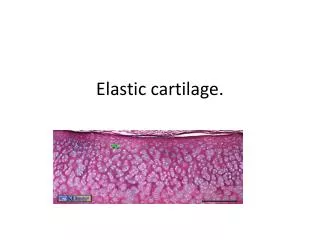
Elastic cartilage ★ is distinguished by the present of elastin in the cartilage matrix. Photomicrograph of elastic cartilage, stained for elastic fibers. Cells are not stained. Resorcin stain.
Elastic cartilage ★ is found in the external ear, the walls of the external auditory canals, the auditory tubes, the epiglottis,and the cuneiform cartilage in the larynx. ★ is surrounded by a perichondrium.
Fibrocartilage ★is a combination of dense connective tissue and hyaline cartilage. ★ is found in intervertebral discs, in attachments of certain ligaments to the cartilaginous surface of bones, and in the symphysis pubis. ★ no perichondrium.
Fibrocartilage ★ Chondrocytes, either singly or in isogenous groups, are usually arranged in long rows separated by coarse collagen type I fibers. ★Numerous collagen fibers either form irregular bundles between the groups of chondrocytes or are aligned in a parallel arrangement along the columns of chondrocytes. Picrosirius-hematoxylin stain
· Histology A: The mesenchyme is the precursor tissue of all types of cartilage. B: Mitotic proliferation of mesenchymal cells gives rise to a highly cellular tissue. C: Chondroblasts are separated from one another by the formation of a great amount of matrix. D: Multiplication of cartilage cells gives rise to isogenous groups, each surrounded by a condensation of territorial (capsular) matrix.
· Growth Interstitial growth: ‘growth from within’ • mitotic division of existing chondrocytes and production of matrix Appositional growth: ‘growth from the surface’ differentiation of new chondrocytes from perichondrial cells and production of matrix at surface
· Repair Poor regeneration of cartilage tissue. Except in young children, damaged cartilage regenerates with difficulty and often incompletely, by activity of the perichondrium, which invades the injured area and generates new cartilage. In extensively damaged areas—and occasionally in small areas—the perichondrium produces a scar of dense connective tissue instead of forming new cartilage.
Medical application Degenerative changes Hyaline cartilage is more susceptible to degenerative aging processes. Calcification of the matrix, preceded by an increase in the size and volume of the chondrocytes and followed by their death, is a common process in some cartilage. Asbestiform degeneration, frequent in aged cartilage, is due to the formation of localized aggregates of thick, abnormal collagen fibrils.
Summary Functions of cartilage Classification of cartilage Matrix of hyaline cartilage Chondrocyte of hyaline cartilage Histology, Growth, and Repair of Hayline cartilage
Homework: 1. What’s the function of cartilage? 2. Classification of cartilage. 3. Review the structure of cartilage.