Download
1 / 36
470 likes | 1.04k Vues
Neonatal Encephalopathy. Julie Parsons, M.D Assistant Professor, Pediatric Neurology November 19, 2009. Disclosures. PTC Therapeutics PTC 124 Clinical Trial for Duchenne Muscular Dystrophy. Objectives. To promote understanding between legal and medical professionals

E N D
NeonatalEncephalopathy Julie Parsons, M.D Assistant Professor, Pediatric Neurology November 19, 2009
Disclosures • PTC Therapeutics • PTC 124 Clinical Trial for Duchenne Muscular Dystrophy
Objectives • To promote understanding between legal and medical professionals • To explain the role of a pediatric neurologist • To define neonatal encephalopathy • To define cerebral palsy • To explore the relationship between neonatal encephalopathy and cerebral palsy
Pediatric Neurologist • A pediatric subspecialist whose expertise is diagnosing and treating disorders in the developing nervous system • This requires completion of a five year residency training program • Board Certification through the American Board of Psychiatry and Neurology • Special Qualification in Child Neurology
The Top 10 Neurologic Conditions • 1. Attention Deficit Hyperactivity Disorder • 2. Seizures and epilepsy • 3. Developmental Delay • 4. Headache • 5. Newborn disorder • 6. Mental Retardation • 7. Macrocephaly/Microcephaly • 8. Motor disturbance • 9. Central Nervous system infection • 10. Neuromuscular Disturbance
Neonatal Encephalopathy • A depression in mental status or alteration of consciousness • Seizures • Abnormal muscle tone and reflexes • Abnormal respiratory function
Risk Factors for Neonatal Encephalopathy • Increased maternal age • Family history of neurologic problems • Maternal thyroid disease or other autoimmune disorder • Coagulopathy or thrombotic disorders • Maternal hypertension • Vaginal bleeding • Maternal infection • Infertility • Intrauterine growth restriction
Sarnat Staging Sarnat and Sarnat, 1976
Correlation of Severity of Clinical Abnormalities and Outcome Sarnat and Sarnat 1976; Volpe 1995
Sarnat and Sarnat Staging • Stage 3 • More than 7 days at Stage 2 • Both associated with poor neurologic prognosis
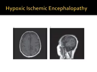
Hypoxic Ischemic Encephalopathy • Hypoxia= Lack of Oxygen • Ischemia= Lack of Perfusion • Hypoxic Ischemic Encephalopathy is caused by a combination resulting in a decreased supply of oxygen to cerebral tissue
Hypoxic Ischemic Encephalopathy • Recognizable Pattern of Progression • 1st 12 hours—bilateral hemispheric involvement leads to decreased level of consciousness • 12 to 24 hours—”apparent improvement” in level of consciousness, but may have seizures which are often refractory • 24 to 72 hours—worsening brainstem dysfunction with altered eye movements, abnormal pupillary response, apnea, bulbar dysfunction (may result in death) • Persistent dysfunction may indicate thalamic or basal ganglia involvement which usually predicts poor prognosis
Diagnosis of HIE: History • Complications of pregnancy, labor, delivery • Placenta previa, cord prolapse, placental abruption, maternal shock • Presence of meconium • Placental condition • chorioamnionitis • Apgar scores (<3 at 5 minutes) • Acid Base status (cord pH <7, BE -12) • Evidence of multiorgan system involvement • Kidney, liver, heart
Differential Diagnosis: Neonatal Encephalopathy • Sepsis • Meningitis • Sedation • Neuromuscular Disease • Trauma • Metabolic
Sepsis • Common antecedent to low Apgar scores and depression • Hypotension • Seizures • Meconium Aspiration • Chorioamnionitis • Bacterial Infections—Meningitis or Group B Strep
Sepsis • Inflammation • Release of cytokines • Widespread endothelial injury • Coagulopathies • Germinal matrix injury • Intraventricular hemorrhage • Stroke
Metabolic Disorders • Biochemical Derangements • Hypoglycemia • Hypocalcemia • Hyponatremia • Hyperammonemia • Acidosis
Metabolic Disorders • Inborn Errors of Metabolism • Symptoms after hours or days of appearing normal • Prominent unexplained vomiting • Hyperpnea in absence of lung disease • Unusual Odor • Family history , or excess fetal loss • Physical Exam findings: cataracts, hepatosplenomegaly
Diagnosis of HIE: EEG • Characteristic Sequence • Initially marked suppression of amplitude and slowing • 24 to 48 hours discontinuous pattern of burst suppression • May deteriorate to isoelectric • Rapid resolution of abnormalities favors good prognosis
Diagnosis of HIE: Imaging • Cranial Ultrasound—increased echogenicity and effacement of sulci although 50% read as normal • Computerized Tomography (CT) • 2 to 5 days maximal extent of parenchymal hypodensities • Atrophy or multicystic encephalomalacia develops later
Diagnosis of HIE: MRI • T2 prolongation 12 to 18 hours-transient edema • T1 high signal after 3 days • T2 shortening after 6 to 7 days denotes permanent injury • DWI (diffusion weighted imaging) more sensitive especially in signaling injury to the thalami and basal ganglia. Maximum sensitivity at 2 to 4 days
Neuropathology • Parasagittal Injury (>34 wks)– spastic quadriplegia • Periventricular White Matter in preterm—spastic diplegia • Basal Ganglia—acute, near total ischemia associated with poor neurologic outcome chorioathetosis and feeding difficulty • Focal/Multifocal– reflects the area of injury • Selective Neuronal Necrosis—HIE
Cerebral Palsy • A chronic neuromuscular disability of central nervous system origin, characterized by abnormal control of movement or posture appearing early in life and not the result of a progressive disease • Intellectual , sensory or behavior problems may accompany but are not a part of the diagnosis • Only 10-15% of children with CP have a history of severe hypoxic ischemic encephalopathy (NCPP)
Diagnosis of Cerebral Palsy • Delayed Milestones • Persistence of developmental reflexes • Pathologic Reflexes • Failure to develop maturational reflexes • No progression or loss of skills by history
Type of Cerebral Palsy • Spastic • Choreoathetotic • Ataxic • Dystonic • Ballismic • Mixed
Clinical Syndromes Spastic Quadriplegia is the form of CP most commonly associated with Hypoxic Ischemic Encephalopathy Spastic Hemiparesis is associated with stroke Athetosis is associated with kernicterus About 30% of children with CP have a brain malformation or cortical dysgenesis
Association of Neonatal Encephalopathy and CP • In <10% of children with CP or MR was there an association with intrapartum or perinatal asphyxia • 75% of children with CP had normal Apgar scores • Apgar < 3 at 15 min 36% with CP • at 20 min 57% with CP • Persistently low Apgar scores at 10, 15, 20 min are associated with poor neurologic outcome
Criteria for Exposure • Birth Asphyxia • Clinical history • Clinical Markers • Abnormal fetal heart rate • Meconium • Laboratory Markers • Fetal Acidosis (cord pH) • Newborn Status • Apgars • Encephalopathy
Conclusions • ACOG guidelines provide a reasonable framework • There is an association between HIE and CP • Neonatal Encephalopathy is a non specific clinical syndrome • Neonatal Encephalopathy must be documented to consider HIE as causation for CP • Continued scientific studies are needed to further understand etiologies of CP