Download
1 / 54
550 likes | 800 Vues
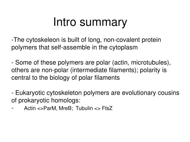
The cytoskeleon is built of long, non-covalent protein polymers that self-assemble in the cytoplasm Some of these polymers are polar (actin, microtubules), others are non-polar (intermediate filaments); polarity is central to the biology of polar filaments

E N D
The cytoskeleon is built of long, non-covalent protein polymers that self-assemble in the cytoplasm • Some of these polymers are polar (actin, microtubules), others are non-polar (intermediate filaments); polarity is central to the biology of polar filaments • Eukaryotic cytoskeleton polymers are evolutionary cousins of prokaryotic homologs: • Actin <>ParM, MreB; Tubulin <> FtsZ Intro summary
CB201.2Intermediate filaments and Polymerization dynamics • Dynamic vs. non-dynamic filaments • Intermediate filaments and nuclear lamins • Measuring polymerization dynamics • NTP hydrolysis during polymerization: treadmilling and dynamic instability • Proteins and drugs that modulate polymerization dynamics
NDP + Pi NTP Polymerization dynamics Actin, ParM, MreB, Tubulin, FtsZ Weak affinity of polymer for monomer (~M) Fast, unidirectional turnover cycle powered by NTP hydrolysis during polymerization
NDP + Pi NTP Polymerization dynamics Actin, ParM, MreB, Tubulin, FtsZ Weak affinity of polymer for monomer (~M) Fast, unidirectional turnover cycle powered by NTP hydrolysis during polymerization Key concept Subunits only come on and off at ends. This allows nm- and msec-scale biochemistry at ends to control the behavior of polymers that exhibit mm- and sec- or min-scale biology
Are all protein polymers controlled by reactions at their ends?Is all the polymerization/depolymerization biochemistry of microtubules and actin confined to their ends?
NDP + Pi NTP Polymerization dynamics Actin, ParM, MreB, Tubulin, FtsZ Weak affinity of polymer for monomer (~M) Fast, unidirectional turnover cycle powered by NTP hydrolysis during polymerization Intermediate filaments High affinity of polymer for monomer (~nM-pM) Polymerization ~irreversible, may occur co-translationally Subsequent dynamics requires protein modification (phosphorylation, proteolysis)
Intermediate filaments; keratin, vimentin, neurofilaments, other cell-type specific filaments, nuclear lamins Mechanical integrity. Nuclear organization (nuclear lamins)
Keratin filaments in an epithelial cell monolayer cultured on glass
Intermediate filament structure IF polypeptide forms an -helix 2 -helices dimerize into a coiled-coil 2 coiled-coils assemble into an anti-parallel tetramer Tetrameric subunits assemble into a non-polar polymer Polymers bundle to give rope-like intermediate filaments
What kind of interaction provides the main driving force that makes two alpha helices interact to form a coiled-coil? • Van der Waals interactions • Electrostatic interactions • Hydrogen bonds • Hydrophobic interactions • All the above
The two polypeptides in a coiled coils can run: • Parallel • Anti-parallel • Either is possible
Keratin mutations compromise the physical integrity of skin Keratin filaments are abundant in skin keratinocytes, where they provide mechanical integrity to the epidermis. Point mutations in skin keratin subunits cause inherited skin disease in humans and mouse models. Severity of the disease correlates with the degree to which polymerization of the mutant keratin subunit into intermediate filaments is inhibited. In these conditions, called epidermolysis bullosa, the epidermis can separate from the dermis, causing severe blistering. Blistering in the mouse models is first evident at sites where the skin experiences the most mechanical stress. Fuchs and Cleveland 1998 Science. 279:514-9
Tissue specific expression of IFs • One of the most striking aspects of IF biology • Keratins in epithelia, GFAP in Glia, Desmin in muscle etc. • ~20 different keratins, always co-expressed as pairs • Useful for histopathology • Keratins in tumor diagnostics • Nestin as a neuronal stem cell marker • What are possible significances of tissue specific expression of IF genes?
Nuclear lamins Nuclear lamina • Lamins are a special type of intermediate filament protein that polymerizes into the nuclear lamina that underlies the nuclear envelope membranes. • Lamin polypeptides contain a nuclear localization sequence (NLS) that makes them enter the nucleus through nuclear pores.
Nuclear lamins depolymerize during mitosis Cdc2.CyclinB kinase phosphatases • The lamina breaks down during mitosis in higher animal cells after phosphorylation by Cdc2.CyclinB kinase, and other kinases • It reforms in the daughter cells through the action of phosphatases
Pi Pi Pi Pi Pi Pi IF dynamics driven by reversible phosphorylation? Binding protein Kinase-X ATP Pi Phosphatase-Y • Phosphorylation promotes lamin depolymerization in mitosis • - IF dynamics are still poorly understood • Keratins in skin and hair become covalently cross-linked as the epithelial cell undergoes terminal differentiation • some IFs more dynamic than others?? e.g. vimentin (mesenchymal cells) more dynamic than keratin (epithelial cells); EMT marker
Nuclear Lamins The nuclear lamina contains 3 types of lamins, A,B and C. All are homologous to intermediate filament subnits and assemble into coiled-coil oligomers Lamin B is prenylated and binds directly to the nuclear envelope membrane. Nucleii in early embryos contain only this lamin type Lamin A and C are generated from the same precursor protein by a complex set of modification at the protein level. Mutation in human lamin-A cause “laminopathies” (Gruenbaum et al 2005 Nat Rev Cell Biol 6:21, Capell and Collins 2006 Nat Rev Genet. 7:940)
Mutations in the Lamin-A gene cause laminopathies AD-EMD, AE-AMD, LGMD1B: muscular dystrophies DCM1A: cardiomyopathy FPLD, GLD: liopdystrophies AWS, HGPS: progerias (premature aging) Worman HJ, Ostlund C, Wang Y. Cold Spring Harb Perspect Biol. 2010 2:a000760. Review
How could two different mutations in the same amino acid in lamin A cause two very different diseases?
NDP + Pi NTP Polymerization dynamics Actin, ParM, tubulin, FtsZ. Weak affinity of polymer for monomer (~M) Polymerization-depolymerization coupled to energy transduction Spontaneous dynamics powered by NTP hydrolysis Intermediate filaments High affinity of polymer for monomer (~nM) Polymerization ~irreversible, occurs at or near ribosome Subsequent dynamics requires protein modification (phosphorylation, proteolysis)
Polymerization dynamics Experiment. Take a solution of a protein that can polymerize and change the conditions to promote polymerization. Then measure [polymer] over time. - Tubulin is ~stably dimeric at 0o with GTP present -->Warm to 37o to polymerize - FtsZ is stably monomeric in GDP -->Add GTP to polymerize - Actin is stably monomeric at very low ionic strength with ATP present. -->Add physiological Mg++ and K+ to polymerize
Measuring tubulin polymerization by light scattering When a light beam is passed through a solution of particles, some of the light is scattered. Scattering increases with the molecular weight Scattering can be quantified by measuring the decrease in light passing through the sample using a spectrophotometer, or by the increase of light emitted at right angles in a fluorimeter. For long polymers like microtubules, the amount of light scattered is proportional to the polymer mass.
SH Measuring actin polymerization by fluorescence spectroscopy using pyrene-actin as probe pH 8 Actin Pyrene-iodoacetate Pyrene- actin Monomer: pyrene quenched by water Low fluorescence Polymer: pyrene buried. High fluorescence
Polymerization dynamics Polymer mass Time (seconds-minutes)
Bulk polymerization dynamics steady state: Polymerization = depolymerization nucleation “lag phase” elongation Polymer mass Time (seconds-minutes)
Bulk polymerization dynamics steady state: Polymerization = depolymerization nucleation “lag phase” elongation Polymer mass Time (seconds-minutes) What is the difference between “steady-state” and “equilibrium”? How would you tell which applied in a case like the graph above?
Describing elongation dynamics c = monomer concentration kon c + koff Assumes: 1. monomors are added and lost only at filament ends at single, unique sites for addition and loss 2. kon and koffare constants that do not change with filament length or polymerization/depolymerization rate
Describing elongation dynamics c = monomer concentration kon c + koff Assumes: 1. monomors are added and lost only at filament ends at single, unique sites for addition and loss 2. kon and koffare constants that do not change with filament length or polymerization/depolymerization rate kon may be “diffusion limited”, this is, it can occur as fast as monomers can collide with the end of the filament What is a typcial value for a diffusion-limited rate constant for proteins?
koff kon Describing polymer dynamics c = monomer concentration kon c + koff Growth rate (J) = kon c - koff At equilibrium, J = 0. kon c = koff Cc = “critical concentration” Cc = Oosawa and Asakura (1975) The thermodynamics of protein polymerization. Acadmeic Press
koff kon Describing polymer dynamics c = monomer concentration kon c + koff Growth rate (J) = kon c - koff At equilibrium, J = 0. kon c = koff Cc = kon J c koff Equilibrium point. Cc = “critical concentration”
A polar polymer without NTP hydrolysis(eg bacterial flagellin) add from minus end add from plus end k+on c k-on c + + k+off k-off
k+on = k-on k-off k+off k-on k+on = A polar polymer without NTP hydrolysis(eg bacterial flagellin) add from minus end add from plus end k+on c k-on c + + k+off k-off If there is a conformational change, then in general But, because DG is the same by either pathway, Cc+ = Cc-
k+on = k-on c k-off k+off k-on k+on = A polar polymer without NTP hydrolysis(eg bacterial flagellin) add from minus end add from plus end k+on c k-on c + + k+off k-off If there is a conformational change, then in general But, because DG is the same by either pathway, plus end The critical concentration must be the same at each end. Depending on c, both ends may either grow, shrink or remain static. J minus end k-off c Cc+ = Cc- k+off
What happens if a microtubule can elongate at multiple sites? kon c + Growth rate (J) = kon c – koff koff ? + • kon is likely to change • koff likely to change • Nothing will change
What happens if a microtubule can elongate at multiple sites? According to a recent paper, multiple independent elongations sites at a microtubule ends cause the off rate increases with tubulin concentration(Gardner, Odde et al 2012 Cell 146:582) In their model, increased growth rate leads to a more irregular microtubule ends, which have higher average off rates and more fluctuations
ATP ATP ADP ADP ADP ADP ADP ADP ADP ADP ADP ADP ADP ADP ADP ADP ADP ADP ADP ADP ADP Nucleotide hydrolysis during polymerization Actin binds ATP and hydrolyzes it during polymerization Phosphate Subunit addition Hydrolysis
ATP ATP ADP ADP ADP ADP ADP ADP ADP ADP ADP ADP ADP ADP ADP ADP ADP ADP ADP ADP ADP Nucleotide hydrolysis during polymerization Actin binds ATP and hydrolyzes it during polymerization Phosphate Subunit addition Hydrolysis ParM, MreB are similar Tubulin binds GTP and hydrolyzes it during polymerization GDP-tubulin GTP-tubulin - + FtsZ, TubZ are biochemically similar but they do not form tubular polymers
Tubulin is a heterodimer of a and b polypeptides(FtsZ and other bacterial tubulin subunit are monomers) Tubulin heterodimers never dissociate after folding -tubulin monomer; rapid GTP exchange, no hydrolysis polymer; No exchange, rapid hydrolysis -tubulin No exchange, no hydrolysis; GTP has purely structural role How do you think this was shown – that the GTP on a-tubulin is structural?
ATP hydrolysis and actin polymerization 100 means 20mol actin monomer has polymerized * Carlier, Pantaloni and Korn, JCB 1984 • Note the kinetic lag between hydrolysis and polymerization. • From this we can infer: • There can be at most a single molecule of ATP-actin at the tip of a polymerizing filament • There can be many ATP actin molecules at the tip
ATP hydrolysis and actin polymerization 100 light scattering units means 20mol actin monomer has polymerized * Carlier, Pantaloni and Korn, JCB 1984 • Note the kinetic lag between hydrolysis and polymerization. • From this we can infer: • Subunit addition to a polymerizing tip requires ATP hydrolysis • Subunit addition does not require ATP hydrolysis
ATP ATP ATP ATP ATP ATP ATP ADP ADP ADP ADP ADP ADP ADP ADP ADP ADP ADP ADP ADP ADP ADP ADP ADP ADP ADP ADP ADP Nucleotide hydrolysis during polymerization Slow polymerization Fast polymerization A kinetic lag between subunit addition and ATP hydrolysis can generate a “cap” of unhydrolyzed subunits
ATP ATP ATP ATP ATP ATP ATP ATP ATP ATP ATP ADP ADP ADP ADP ADP ADP ADP ADP ADP ADP ADP Nucleotide hydrolysis “weakens” the polymer For both actin filaments and microtubules, ATP (GTP) hydrolysis has the effect of increasing the dissociation rate constant Slow Fast
ATP ATP ATP ATP ATP ATP ATP ATP ATP ATP ATP ADP ADP ADP ADP ADP ADP ADP ADP ADP ADP ADP Nucleotide hydrolysis “weakens” the polymer For both actin filaments and microtubules, ATP (GTP) hydrolysis has the effect of increasing the dissociation rate constant Slow Fast ATP AMP-PNP To do this experiment, you need either a mutation in the actin, or a non-hydrolyzeable ATP analog
ATP ATP ATP ADP ADP ADP ADP ADP ADP ADP ADP ADP ADP ADP ADP Treadmilling of pure actin driven by ATP hydrolysis Pi Slow ADP ATP Note. Treadmilling of pure actin is very slow, and it is not clear if this reaction is relevant inside cells, where turnover is often very fast
Treadmilling occurs at steady state because the critical concentration is lower on the barbed than the pointed end Barbed end J Cc+ Cc- Pointed end k-off c k+off Steady state monomer concentration, where growth on plus ends is exactly balanced by shrinkage of minus ends
ADP.Pi ADP.Pi ADP.Pi ADP.Pi ADP.Pi ATP ATP ATP ADP ADP ADP ADP ADP ADP ADP Two small proteins accelerate actin dynamics in cells Pi Cofilin Profilin ADP ATP • - Cofilin only binds ADP-actin. It greatly weakens the filament, promoting faster dissociation of subunits from ends, as well as severing • Profilin helps recycle actin monomer • Cofilin and profilin conserved throughout eukaryotes • The exact pathway of depolymerization in cells is still unknown
Microtubules: dynamic instability Microtubules alternate between bouts of polymerization and depolymerization. This is true for pure tubulin in vitro, and also in cytoplasm, though the rates differ. Rhodamine-tubulin polymerizing from a centrosome in frog egg extract. Observation by widefield fluorescence microscopy
Dynamic Instability is driven by GTP hydrolysis Pi Growing microtubule GTP-tubulin GDP Nucleotide exchange Catastrophe Rescue GTP Shrinking microtubule GDP-tubulin Model: GTP subunits like to be straight, while GDP subunits like to be curved. GTP hydrolysis puts the lattice under stress. Growing microtubules are stabilized by a cap of GTP-subunits. When this cap is lost the microtubule depolymerizes rapidly by a kind of tearing or peeling apart. (Nogales and Wang 2006 Curr Opin Struct Biol 16:221-9)
How long is the GTP cap? Pi Growing microtubule GTP-tubulin Short cap? Long cap? How would you ask this question experimentally?
Cellular factors regulate Dynamic Instability MAPs stabilize microtubules Kinesin-13 promotes catastrophes Pi GDP GTP