Download
1 / 52
560 likes | 1.88k Vues
Pentose phosphate pathway and NADPH. UNIT II: Intermediary Metabolism. Figure 13.1. Hexose monophosphate pathway shown as a component of the metabolic map. Overview. The pentose phosphate pathway (a.k.a, hexose monophosphate shunt, or 6-phosphogluconate pathway) occurs in cytosol of the cell

E N D
Pentose phosphate pathway and NADPH UNIT II: Intermediary Metabolism
Figure 13.1. Hexose monophosphate pathway shown as a component of the metabolic map
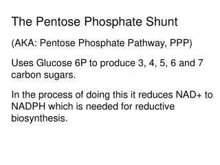
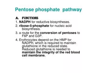
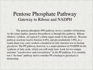
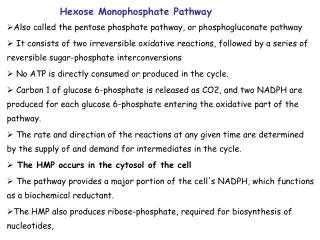
Overview • The pentose phosphate pathway (a.k.a, hexose monophosphate shunt, or 6-phosphogluconate pathway) occurs in cytosol of the cell • It consists of two, irreversible oxidative reactions, followed by a series of reversible sugar-phosphate interconversions. • No ATP is directly consumed or produced in the cycle. Carbon one of G-6-P is released as CO2, & 2 NADPH are produced for each G-6-P molecule entering the oxidative part of the pathway. • The rate & direction of the reversible reactions of the pathway are determined by the supply of & demand for intermediates of the cycle. • The pathway provides a major portion of the body’s NADPH, which functions as a biochemical reductant. • It also produces ribose-5-P required for biosynthesis of nucleotides, & provides a mechanism for metabolic use of five-carbon sugars obtained from diet or degradation of structural CHOs in the body.
II. Irreversible oxidative reactions • The oxidative portion of pentose phosphate pathway consists of 3 reactions that lead to formation of ribulose 5-P, CO2, & 2 molecules of NADPH for each molecule of G-6-P oxidized. • This portion of the pathway is particularly important in the liver & lactating mammary glands, which are active in the biosynthesis of fatty acids, in adrenal cortex, which is active in the NADPH-dependent synthesis of steroids, & in RBCs, which require NADPH to keep glutathione reduced
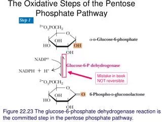
A. Dehyerogenation of glucose-6-phosphate • Glucose 6-phosphate dehydrogenase (G6PD) catalyzes an irreversible oxidation of G-6-P to 6-phosphogluconolactone in a reaction that is specific for NADP+ as its coenz. • The pentose phosphate pathway is regulated primarily at the G6PD reaction. NADPH is a potent competitive inhibitor of the enz, &, under metabolic conditions, the ratio of NADPH/NADP+ is sufficiently high to substantially inhibit enz activity. • However, with increased demand for NADPH, the ratio of NADPH/NADP+ decreases & flux through the cycle increases in response to the enhanced activity of G6PD. • Insulin enhances G6PD gene expression, & flux through the pathway increases in the well-fed state
B. Formation of ribulose-5-phosphate • 6-phosphogluconolactone is hydrolyzed by 6-phosphogluconolactone hydrolase. The reaction is irreversible & not rate-limiting. • The subsequent oxidative decarboxylation of 6-phosphogluconate is catalyzed by 6-phosphogluconate dehydrogenase. This irreversible reaction produces a pentose sugar-phosphate (ribulose 5-P), CO2 (from C-1 of glucose), & a 2nd molecule of NADPH.
Figure 13.2. Reactions of the hexose monophosphate pathway. Enzymes numbered above are 1) glucose 6-phosphate dehydrogenase and 6-phosphogluconolactone hydrolase, 2) 6-phosphogluconate dehydrogenase, 3) ribose 5-phosphate isomerase, 4) phosphopentose epimerase, 5) and 7) transketolase (coenzyme: thiamine pyrophosphate), and 6) transaldolase.
III. Reversible nonoxidative reactions • The nonoxidative reactions of pentose phosphate pathway occur in all cell types synthesizing nucleotides & nucleic acids. • These reactions catalyze the interconversion of 3-, 4-, 5-, 6-, & 7-carbon sugars. These reversible reactions permit ribulose-5-P (produced by oxidative portion of pathway) to be converted either to ribose 5-P (needed for nucleotide synthesis) or to intermediates of glycolysis, F-6-P & glyceraldehyde 3-P.
E.g., many cells that carry out reductive biosynthetic reactions have a greater need for NADPH than for ribose-5-P. In this case, transketolase (which transfers 2-C units) & transaldolase (which transfers 3-C units) convert ribulose 5-P produced as an end-product of the oxidative reactions to glyceraldehyde 3-P & F-6-P, which are intermediates of glycolysis. • In contrast, under conditions in which the demand for ribose for incorporation into nucleotides & nucleic acids is greater than the need for NADPH, the nonoxidative reactions can provide the biosynthesis of ribose-5-P from glyceraldehyde 3-P & F-6-P in the absence of the oxidative steps
Figure 13.3. Formation of ribose 5-phosphate from intermediates of glycolysis.
IV. Uses of NADPH • The coenz NADP+ differs from NAD+ only by the presence of a P-group (-PO4=) on one of the ribose units. • This seemingly small change in structure allows NADP+ to interact with NADP+-specific enz’s that have unique roles in the cell. E.g., the steady-state ratio of NADP+/NADPH in the cytosol of hepatocytes is ~ 0.1, which favors the use of NADPH in reductive biosynthetic reactions. • This contrasts with the high ratio of NAD+/NADH (~ 1000 in the cytosol of hepatocytes), which favors an oxidative role of NAD+.
A. Reductive biosynthesis • NADPH can be thought of as a high-energy molecule, much in the same way as NADH. However, the e’s of NADPH are destined for use in reductive biosynthesis, rather than for transfer to oxygen as is the case with NADH. - Thus, in the metabolic transformations of the pentose phosphate pathway, part of the energy of G-6-P is conserved in NADPH, a molecule that can be used in reactions requiring a high electron-potential e-donor.
B. Reduction of hydrogen peroxide • Hydrogen peroxide is one of a family of reactive oxygen species that are formed from the partial reduction of molecular oxygen. • These cpds are formed continuously as by-products of aerobic metabolism, through reactions with drugs & environmental toxins, or when level of antioxidants is diminished, all creating the condition “oxidative stress”. • The highly reactive oxygen intermediates can cause serious chemical damage to DNA, proteins, & unsaturated lipids, and can lead to cell death. • These reactive oxygen species have been implicated in a number of pathologic processes including reperfusion injury, cancer, inflammatory disease, and aging. • The cell has several protective mechanisms that minimize the toxic potential of these cpds.
1. Enzymes that catalyze antioxidant reactions: - A tripeptide-thiol (γ-glutamylcysteinylglycine) present in most cells, can chemically detoxify hydrogen peroxide • This reaction, catalyzed by the selenium-requiring glutathione peroxidase, forms oxidized glutathione, which no longer has protective property • The cell regenerates reduced glutathione in a reaction catalyzed by glutathione reductase, using NADPH as a source of reducing electrons • Thus, NADPH indirectly provides e’s for the reduction of hydrogen peroxide • Additional enz’s, such as superoxide dismutase & catalase, catalyze the conversion of other toxic oxygen intermediates to harmless products • As a group, these enz’s serve as a defense system to guard against the toxic effects of reactive oxygen species
Figure 13.5 A. Formation of reactive intermediates from molecular oxygen. B. Actions of antioxidant enzymes. G-SH = reduced glutathione; G-S-S-G = oxidized glutathione.
Figure 13.6. A. Structure of glutathione (G-SH). [Note: Glutamate is linked to cysteine through a γ-carboxyl, rather than an α-carboxyl.] B. Glutathione-mediated reduction of hydrogen peroxide by NADPH.
Note: - RBCs are totally dependent on the pentose phosphate pathway for their supply of NADPH because, unlike other cell types, RBCs do not have an alternate source for this essential coenz. • If G6PD is compromised in some way, NADPH levels will fall, & oxidized glutathione can’t be reduced • As a result, hydrogen peroxide will accumulate, threatening memb stability & causing RBC lysis
2. Antioxidant chemicals • A number of intracellular reducing agents such as ascorbate, vitamin E, & β-carotene, are able to reduce &, thus, detoxify oxygen intermediates in the lab. • Consumption of foods rich in these antioxidant cpds has been correlated with a reduced risk for certain types of cancers, as well as decreased frequency of certain other chronic health problems • Thus, it is tempting to speculate that the effects of these cpds are, in part, an expression of their ability to quench the toxic effect of oxygen intermediates • However, clinical trials with antioxidants as dietary supplements have failed to show clear beneficial effects. • In the case of dietary supplementation with β-carotene, the rate of lung cancer in smokers increased rather than decreased. • Thus, the health-promoting effects of dietary fruits & vegetables probably reflects a complex interaction among many naturally occurring cpds, which has not been duplicated by consumption of isolated antioxidant cpds
C. Cytochrome P450 monooxygenase system • Monooxygenases (mixed function oxidases) incorporate one atom from molecular oxygen into a substrate (creating a hydroxyl group), with the other atom being reduced to water • In the cytochrome P450 monooxygenase system, NADPH provides the reducing equivalents required by this series of reactions • This system performs different functions in two separate locations in cells. The overall reaction catalyzed by Cyt-P450 enz is: R-H + O2 + NADPH + H+ → R-OH + H2O + NADP+ Where R may be a steroid, drug, or other chemical Note: Cyt-P450s (CYPs) are actually a superfamily comprised of 100s of genes, coding for related, heme-containing enz’s that participate in a broad variety of reactions
Figure 13.7 Cytochrome P450 monooxygenase cycle.
1. Mitochondrial system: • The function of the mitoch Cyt-P450 monooxygenase system is to participate in the hydroxylation of steroids, a process that makes these hydrophobic cpds more water soluble • E.g., in the steroid hormone-producing tissues, e.g., placenta, ovaries, testes, & adrenal cortex, it is used to hydroxylate intermediates in the conversion of cholesterol to steroid hormones • The liver uses this system in bile acid synthesis, & the kidney uses it to hydroxylate vitamin 25-hydroxycholecalciferol (vitamin D) to its biologically active 1,25-hydroxylated form
2. Microsomal system: • An extremely important function of the microsomal Cyt-P450 monooxygenase system found associated with memb’s of sER (particularly in liver) is the detoxification foreign cpds (xenobiotics) • Xenobiotics include numerous drugs & such varied pollutants as petroleum products, carcinogens, & pesticides • The Cyt-P450 monooxygenase system can be used to hydroxylate these toxins, again using NADPH as source of reducing equivalents • The purpose of these modifications is 2-fold. 1st, it may itself activate or inactivate a drug or 2nd, make a toxic cpd more soluble, thus facilitating its excretion in the urine or feces. • Frequently, however, the new hydroxyl group will serve as a site for conjugation with a polar cpd, such as glucuronic acid, which will significantly increase the cpd’s solubility
D. Phagocytosis by white blood cells • Phagocytosis is the ingestion by receptor-mediated endocytosis of m/o’s, foreign particles, & cellular debris by cells such as neutrophils & macrophages (monocytes) • It is an important body defense mechanism, particularly in bacterial infections. • Neutrophils & monocytes are armed with both oxygen-independent & oxygen-dependent mechanisms for killing bacteria. • The oxygen-dependent mechanisms include the myeloperoxidase (MPO) system & a system that generates oxygen-derived free radicals • Oxygen-independent systems use pH changes in phagolysosomes & lysosomal enz’s to destroy pathogens
Overall, the MPO system is the most potent of the bactericidal mechanisms. • An invading bacterium is recognized by the immune system & attacked by antibodies that bind it to a receptor on a phagocytic cell • After internalization of the m/o has occurred, NADPH oxidase, located in the leukocyte CM, converts molecular oxygen from the surrounding tissue into superoxide • The rapid consumption of molecular oxygen that accompanies formation of superoxide is referred to as the respiratory burst
Note: • NADPH oxidase is a complex enz, with subunits containing a cytochrome & a flavin coenz group • Genetic deficiencies in this enz cause chronic granulomatosis, a disease characterized by severe, persistent, chronic pyogenic infections
Next, superoxide is spontaneously converted into hydrogen peroxide. Any superoxide that escapes the phagolysosome is converted to hydrogen peroxide by superoxide dismutase (SOD). • This product is then neutralized by catalase or glutathione peroxidase • In the presence of MPO, a lysosomal enz present within the phagolysosome, peroxide plus chloride ions are converted into hypochorous acid (HOCl, the major component of household bleach), which kills the bacteria. • Excess peroxide is either neutralized by catalase or by glutathione peroxidase
Figure 13.8 Phagocytosis and the oxygen dependent pathway of microbial killing. IgG = the antibody immunoglobulin G.
E. Synthesis of nitric oxide • Nitric oxide (NO) is recognized as a mediator in a broad array of biologic systems • NO is the endothelium-derived relaxing factor, which causes vasodilation by relaxing vascular smooth muscle. • NO also acts as a neurotransmitter, prevents platelet aggregation, & plays an essential role in macrophage function. • NO has a very short half-life in tissues (3-10 seconds) because it reacts with oxygen & superoxide, & then is converted into nitrates & nitrites. Note: - NO is a free radical gas that is confused with nitrous oxide (N2O), the “laughing gas” that is used as an anesthetic & is chemically stable
1. Synthesis of NO: - Arg, O2, & NADPH are substrates for cytosolic NO synthase. - Flavin mononucleotide (FMN), flavin adenine dinucleotide (FAD), heme, & tetrahydropterin are coenz’s for the enz, & NO & citrulline are products of the reaction • Three NO synthases have been identified. Two are constitutive (synthesized at a constant rate regardless of physiologic demand), Ca2+-calmodulin-dependent enzymes. They are found primarily in endothelium (eNOS), & neural tissue (nNOS), & constantly produce low levels of NO • An inducible, Ca2+-independent enz. (iNOS) can be expressed in many cells, including hepatocytes, macrophages, monocytes, & neutrophils • The specific inducers for NO synthase vary with cell type, & include tumor necrosis factor-α, bacterial endotoxins, & inflammatory cytokines. These cpds have been shown to promote synthesis of iNOS, which can result in large amounts of NO being produced over hours or even days.
Figure 13.9 Synthesis and some of the actions of nitric oxide.
2. Action of NO on vascular endothelium: • NO is an important mediator in the control of vascular smooth muscle tone • NO is synthesized by eNOS in endothelial cells & diffuses to vascular smooth muscle, where it activates the cytosolic form of guanylate cyclase Note: this reaction is analogous to the formation of cAMP by adenylate cyclase, except that this guanylate cyclase is not membrane-associated • The resultant rise in cGMP causes muscle relaxation through activation of protein kinase G, which phospho. myosin light-chain kinase & renders it inactive, thereby decreasing smooth muscle contraction Note: vasodilator nitrates, such as nitroglycerin & nitroprusside, are metabolized to nitric oxide, which causes relaxation of vascular smooth muscle &, therefore, lowers blood pressure. Thus, NO can be envisioned as an endogenous nitrovasodilator
3. Role of NO in mediating macrophage bactericidal activity: - In macrophages, iNOS activity is normally low, but synthesis of the enz is significantly stimulated by bacterial LPS & γ-IFN release in response to infection. • Activated macrophages form superoxide radicals that combine with NO to form intermediates that decompose, forming the highly bactericidal OH•- radical Note: NO production in macrophages is also effective against viral, fungal, helmintic, & protozoan infections 4. Other functions of NO: - NO is a potent inhibitor of platelet aggregation (by activating cGMP pathway). It is characterized as a neurotransmitter in the brain
V. Glucose 6-P dehydrogenase deficiency • G6PD deficiency is an inherited disease characterized by hemolytic anemia caused by the inability to detoxify oxidizing agent • G6PD deficiency is the most common disease-producing enz abnormality in humans, affecting > 200 million individuals worldwide. • This deficiency has the highest prevalence in the Middle East, tropical Africa & Asia, & parts of the Mediterranean • G6PD deficiency is X-linked, & is in fact, a family of deficiencies caused by > 400 different mutations in the gene coding for G6PD. Only some of these mutations cause clinical symptoms
The lifespan of many individuals with G6PD deficiency is somewhat shortened as a result of complications arising from chronic hemolysis • This slightly negative effect of G6PD deficiency has been balanced in evolution by an advantage in survival, an increased resistance to falciparum malaria shown by female carrier of the mutation Note: sickle cell trait & β-thalassemia minor also confer resistance
A. Role of G6PD in RBCs • Diminished G6PD activity impairs the ability of the cell to form NADPH that is essential for the maintenance of reduced glutathione pool. This results in decrease in the cellular detoxification of free radicals & peroxides formed within cell • Glutathione also helps maintain the reduced states of sulfhydryl groups in proteins, including Hb. Oxidation of those sulfhydryl groups leads to formation of denatured proteins that form insoluble masses (called Heinz bodies) that attach to the red cell memb’s. • Additional oxidation of memb proteins causes the red cells to be rigid & non-deformable, & they are removed from the circulation by macrophages in spleen & liver
Figure 13.10. Pathways of G-6-P mtabolism in the erythrocyte
Figure 13.11 Heinz bodies in erythrocytes of patient with G6PD deficiency.
Although G6PD deficiency occurs in all cells of the affected individual, it is most severe in erythrocytes, where the pentose phosphate pathway provides the only means of generating NADPH • Other tissues have alternative sources for NADPH production (e.g., NADP+-dependent malate dehydrogenase) that keep glutathione reduced • The erythrocyte has no nucleus or ribosomes & can’t renew its supply of the enz. Thus, RBCs are particularly vulnerable to enz variants with diminished stability
B. Precipitating factors in G6PD deficiency • Most individuals who have inherited one of the many G6PD mutations do not show clinical manifestations. However, some patients with G6PD deficiency develop hemolytic anemia if they are treated with an oxidant drug, ingest fava beans, or contract a severe infection • Oxidant drugs: commonly used drugs that produce hemolytic anemia in patients with G6PD deficiency are best remembered from the mnemonic AAA = Antibiotics (e.g., sulfa, methoxazole & chloramphenicol), Antimalarials (e.g., primaquine but not quinine), and Antipyretics (e.g., acetanilid but not acetaminophen)
Favism: some forms of G6PD deficiency, e.g., the Mediterranean variant, are particularly susceptible to the hemolytic effect of the fava bean, a dietary staple in Mediterranean region. Favism, the hemolytic effect of ingesting fava beans, is not observed in all individuals with G6PD deficiency, but all patients with favism have G6PD deficiency • Infection: infection is the most common precipitating factor of hemolysis in G6PD deficiency. The inflammatory response to infection results in the generation of free radicals in macrophages, which can diffuse into the RBCs & cause oxidative damage • Neonatal jaundice: babies with G6PD deficiency may experience neonatal jaundice appearing 1-4 days after birth. The jaundice, which may be severe, results from impaired hepatic catabolism of heme or increased production of bilirubin
C. Properties of the variant enzymes • Almost all G6PD variants are caused by point mutations in the G6PD gene. • Some mutations do not disrupt the structure of the enz’s active site &, hence, do not affect enzymic activity • However, many mutant enz’s show altered kinetic properties. E.g., variant enz’s may show decreased catalytic activity, decreased stability, or an alteration of binding affinity for NADP+, NADPH, or G-6-P • Severity of disease usually correlates with amount of residual enz activity in patients’ RBCs. E.g., variants can be classified as:
Figure 13.12 Classification of G6PD deficiency variants.
G6PD A- is the prototype of the moderate (class III) form of disease. The RBCs contain an unstable, but kinetically normal G6PD, with most of the enz activity present in the reticulocytes & younger erythrocytes • The oldest cells, therefore, have the lowest level of enz activity, & are preferentially removed in a hemolytic episode. • G6PD Mediterranean is the prototype of a more severe (class II) deficiency in which the enz shows normal stability but scarcely detectable activity in all RBCs. • Class I mutations are often associated with chronic non-spherocytic anemia, which occurs even in absence of oxidative stress
Figure 13.13 Decline of erythrocyte G6PD activity with cell age for the three most commonly encountered forms of the enzyme.
D. Molecular biology of G6PD • Cloning of G6PD gene & the sequencing of its cDNA have permitted identification of mutations that cause G6PD deficiency • More than 300 different mutations or mutation combinations have been identified in this gene, a finding that explains the numerous biochemical variants • Most of these DNA changes are missense, point mutations. Both G6PD A- & G6PD Mediterranean represent mutant enz’s that differ from the respective normal variants by a single aa • Large deletions or frameshifts mutations have not been identified, suggesting that complete absence of G6PD activity is probably lethal
5 yr old boy presents to the emergency room: febrile, pale, tachycardic, tachypneic and minimally responsive AM: good health PM: abdominal pain, headache, fever by late evening: tachypneic and incoherent Lab tests: massive nonimmune intravasuclar hemolysis and hemoglobinurea The patient is of Greek ethnicity. Mother notes that although there is no family history of hemolysis, she has some European cousins with a ‘blood problem’ She later recalls that her son had been eating fava beans in the garden while she worked in the yard
Summary • The pentose phosphate pathway consists of 2 irreversible oxidative reactions followed by a series of reversible sugar-phosphate interconversions • No ATP is directly consumed or produced in the cycle • The oxidative portion is particularly important in liver & mammary glands, which are active in biosynthesis of fatty acids, in adrenal cortex, which is active in NADPH-dependent synthesis of steroids, & in erythrocytes, which require NADPH to keep glutathione reduced • G-6-P is irreversibly converted to ribulose-5-P, & 2 NADPH are produced. • The regulated step is G6PD, which is strongly inhibited by NADPH • Reversible nonoxidative reactions interconvert sugars. This part of pathway is the source of ribose 5-P required for nt & nucleic acid synthesis • Because reactions are reversible, they can be entered from F-6-P & GA 3P (glycolytic intermediates) if ribose is needed & G6PD is inhibited
NADPH is a source of reducing equivalents in reductive biosynthesis, such as production of fatty acids & steroids. It is also required for reduction of hydrogen peroxide, providing the reducing equivalents required by glutathione (GSH). • GSH is used by glutathione peroxidase to reduce peroxide to water. The oxidized glutathione is reduced by glutathione reductase, using NADPH as the source of e’s • NADPH provides reducing equivalents for cyt-P450 monooxygenase system, which is used in hydroxylation of steroids to produce steroid hormones, bile acid synthesis by liver, & activation of vitamin D. the system also detoxify foreign cpds, e.g., drugs & varied pollutants, including carcinogens, pesticides, & petroleum products
NADPH provides reducing equivalents for phagocytes in the process of eliminating invading m/o’s • NADPH oxidase uses molecular oxygen & NADPH e’s to produce superoxide radicals, which, in turn, can be converted to peroxide, hypochlorous acid, & hydroxyl radicals. Myeloperoxidase is an important enz in this pathway • A genetic defect in NADPH oxidase causes chronic granulomatosis, a disease characterized by severe, persistent, chronic pyogenic infections • NADPH is required for synthesis of nitric oxide (NO), an important molecule that causes vasodilation by relaxing vascular smooth muscle, acts as a kind of neurotransmitter, prevents platelet aggregation, & helps mediate macrophage bactericidal activity