Download

1 / 48
480 likes | 497 Vues
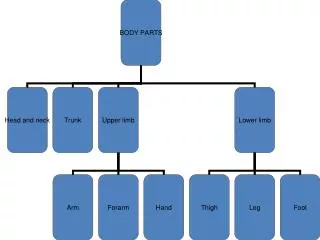
ARTERIES OF UPPER LIMB Dr S. Bhat. ARTERIES OF UPPER LIMB. Axillary artery Brachial artery Radial artery Ulnar artery The arches in hand are Superficial palmar arch Deep palmar arch Dorsal carpal arch Palmar carpal arch. AXILLARY ARTERY.

E N D
ARTERIES OF UPPER LIMB Axillary artery Brachial artery Radial artery Ulnar artery The arches in hand are Superficial palmar arch Deep palmar arch Dorsal carpal arch Palmar carpal arch
AXILLARY ARTERY Continuation of the subclavian artery Extent: • Outer border of the 1st rib to the • Lower border of teres major muscle • Continues as brachial artery • Artery is subdivided into three parts by the pectoralis minor muscle.
First part: It extends from outer border of first rib to the upper border of pectoralis minor Branches: 1. Superior thoracic artery • Arises near lower border of subclavius muscle • Supplies thoracic wall and breast
RELATIONS Laterally: Lateral and posterior cord of brachial plexus Medially: Axillary vein Anterior: • Skin • Superficial fascia • Pectoralis major • Clavipectoral fascia Posterior: • 1st intercostal space • Serratus anterior • Medial cord of brachialplexus
Second part: lies deep to pectoralis minor Branches: 1. Lateral thoracic artery • Related to anterior axillary lymph nodes • Larger in females and supplies branches to breast • Anastomoses with internal thoracic and subscapular arteries
2. Thoracoacromaial artery • Pierces clavipectoral fascia Branches: a. Pectoral - Pectoral muscles and breast b. Deltoid - Pectoralis major and deltoid c. Acromial - Coracoid process anastomoses with deltoid branch of thoraco acromial and suprascapular arteries d. Clavicular- Sternoclavicular joint and subclavius
RELATIONS: Anterior: • Pectoralis major and • Pectoralis minor Posterior: • Posterior cord of brachial plexus • Coracobrachialis Medially: • Medial cord of brachial plexus, • Axillary vein Laterally: • Lateral cord of brachial plexus
Third part: It extends from lower border of pectoralis minor upto the level of lower border of teres major muscle. Branches: a) Anterior circumflex humeral • Lower border of subscapularis • passes deep to Coracobrachialis muscle and biceps, • winds round the surgical neck • Anastomoses with posterior circumflex humeral
b) Posterior Circumflex humeral artery: • Larger than anterior circumflex • Enters quandragular space • Accompanied by axillary nerve • Anastomoses with anterior circumflex around surgical neck of humerus. • also anastomoses with the branches of suprascapular and thoracoacromial arteries
c) Subscapular artery: • Inferior aspect of 3rd part of axillary artery at lower border of subscapularis • Largest branch of axillary artery and supplies latissimus dorsi and serratus anterior. • Reaches inferior angle of scapula and anastomoses with lateral thoracic,intercostal and deep branch of transverse cervical artery. Circumflex scapular artery: • Upper triangular space, infraspinous fossa • Anastomoses around scapula
1ST RIB SKIN SERRATUS ANTERIOR SUPERFICIAL FASCIA 2ND RIB CLAVIPECTORAL FASCIA PECTORALIS MAJOR SUBSCAPULARIS PECTORALIS MINOR TERES MAJOR LATISSIMUS DORSI AXILLARY ARTERY
Teres major muscle Lateral thoracic artery
Thoracoacromial artery Posterior circumflex humeral artery Superior thoracic artery Anterior circumflex humeral artery Lateral thoracic artery Subscapular artery
BRACHIAL ARTERY It is the continuation of the axillary artery Extent: • Lower border of teres major muscle to neck of radius in cubital fossa • Terminal branches: Radial and Ulnar arteries Branches: • Profunda brachii artery • Nutrient artery to humerus • Muscular branches • Superior ulnarcollateral • Inferior ulnarcollateral
Profunda brachii artery: • Largest branch Branches: • Muscular branches-triceps,deltoid • Deltoid branch • Middle collateral • Radial collateral
Nutrient artery: • at the level of Insertion of coracobrachialis muscle Superior ulnar collateral: • Little below middle of arm Inferior ulnar collateral: • 5 cm above elbow Muscular branches: • To muscles of anterior compartment of arm
ULNAR ARTERY: Branches: Forearm: a) Anterior ulnar recurrent artery b) Posterior ulnar recurrent artery c) Common interosseous artery Anterior interosseous artery Posterior interosseous artery d) Muscular branches e) Palmar carpal branch f) Dorsal carpal branch-above pisiform deep to flexor carpi ulnaris
Branches of Ulnar artery in hand • Superficial branch-takes part in the formation of superficial palmar arch by joining with radial artery • Deep branch- passes between hypothenar muscles and anastomoses with terminal part of radial artery-deep palmar arch.
Radial artery Branches: Forearm: • Radial recurrent artery • Muscular branches • Dorsal carpal branch • Palmar carpal branch • Superficial palmar branch
Superficial Palmar branch Given as it winds around lateral side of wrist-ends in superficial palmar arch Dorsal Carpal branch: Given off from radial artery as it passes deep to extensor muscles of the thumb Dorsum of hand: 1st Dorsal metacarpal artery: Before it between two heads of first dorsal interosseous muscle. Divides into two branches to supply adjacent sides of thumb and index finger.
Palm of hand: A)Arteria radialis indicis B) Arteria princeps pollicis C) Main artery taking part in deep palmar arch Arteria Princeps pollicis: As it turns medially into palm of hand. Passes deep to oblique head of adductor pollicis to reach proximal phalnx of thumb. It divides into two branches to supply skin and subcutaneous tissue of the thumb. Main nutrient artery to 1st metacarpal bone.
Arteria Radialis Indicis: Runs between transverse head of adductor pollicis and 1st DI. It supplies radial side of index finger and anastomoses with digital artery supplying medial side of index finger
SUPERFICIAL PALMAR ARCH Situation: In palm of hand infront of flexor tendons,lumbricals,palmar digital branches of median nerve and behind skin and palmar aponeurosis,palmaris brevis Formation: Superficial branch of the ulnar artery joined laterally by a) Superficial branch of radial artery or b) Arteria radialis indicis or c) Arteria Princeps pollicis
Branches: • Palmar digital artery to medial side of little finger b) Three common palmar digital arteries Each common palmar Digital artery Two proper digital arteries Adjacent sides of index,middle ring and little fingers except Radial side of index finger and thumb
All the arteries freely anastomose at finger tips and at the interphalengeal joints by their small branches Two dorsal branches- soft tissue of middle and distal phalnx including nail bed Palmar digital arteries supply-phalanges, metacarpophalengeal and interphalengeal joints They supply mainly digits
PALMAR or VOLAR CARPAL ARCH Situation: Near the lower border of pronator quadratus infront of lower end of radius,ulna and carpal bones Formation: • Palmar carpal branch of ulnar artery • Palmar carpal branch of radial artery • Anterior interosseous artery • Recurrent branch of deep palmar arch Supply articulations of wrist, carpal bones, lower epiphysis of radius and ulna
Dorsal Carpal Arch Situation: Deep to extensor tendons on the posterior surface of lower end of radius Formation: • Dorsal carpal branch of ulnar artery • Dorsal carpal branch of radial artery • Anterior interosseous artery • Posterior interosseous artery Supplies lower epiphyseal parts of radius and Ulna
Branches: Three slender dorsal metacarpal arteries which run on 2nd,3rd,and 4th dorsal interosseoi muscles and divide into dorsal digital branches which supply medial four fingers.They anastomose with palmar digital branches of the superficial palmar arch Proximal perforating arteries-Deep palmar arch Distal perforating arteries Near their bifurcation-Palmar digital branches of superficial palmar arch.
Anterior and posterior interosseous arteries
Deep Palmar Arch: Formation: Deep branch of ulnar artery with terminal part of radial artery Situation: Proximal ends of metacarpal bones,interossei muscles,under cover of adductor pollicis muscle and deep to flexor tendons and lumbricals BRANCHES: • 3 palmar metcarpal arteries • 3 perforating arteries • Recurrent artery
Palmar metacarpal arteries- Interossei of 2nd,3rd and 4th spaces Anastomose with common digital branches of superficial palmar arch at the clefts between the fingers Nutrient branches to medial 4 metacarpal bones Perforating branches Pass dorsally between dorsal interossei muscles and anastomose with dorsal metacarpal arteries. Recurrent branch: Supply carpal bones,joints between carpal bones and wrist joint
ANASTOMSIS AROUND SCAPULA Free anastomosis takes place around scapula between the branches of the subclavian and axillary artery. • Suprascapular artery-thyrocervical trunk • Deep branch of transverse cervical artery • Subscapular artery and its branch-circumflex scapular artery