Download
1 / 54
610 likes | 990 Vues
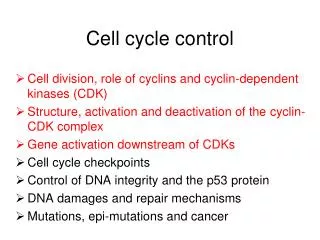
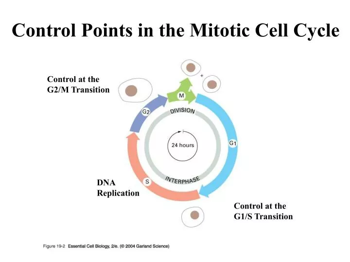
Control Points in the Mitotic Cell Cycle. Control at the G2/M Transition. DNA Replication. Control at the G1/S Transition. Control Points in the Mitotic Cell Cycle. Positive Control by Cyclin/Cdk. Negative Control By p53 (checkpoint). Cyclin Protein Levels Vary

E N D
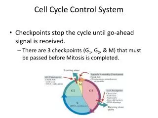
Control Points in the Mitotic Cell Cycle Control at the G2/M Transition DNA Replication Control at the G1/S Transition
Control Points in the Mitotic Cell Cycle Positive Control by Cyclin/Cdk Negative Control By p53 (checkpoint)
Cyclin Protein Levels Vary During the Cell Cycle
Cyclin/Cdk Complexes During the Cell Cycle Expression of M-cyclin Gene Ubiquitin-dependent proteolysis Ubiquitin-dependent proteolysis Expression of S-cyclin Gene
Cyclin/Cdk Phosphorylates Rb Protein (RTK) (Ras-MAP Kinase) Activation of “S-Phase Genes”
Control Points in the Mitotic Cell Cycle Positive Control by Cyclin/Cdk Checkpoint by p53
The p53 Cell Cycle Checkpoint or Short Telomeres (ATM Kinase)
The p53 Cell Cycle Checkpoint or Short Telomeres DNA Repair System (ATM Kinase) Programmed Cell Death
Connection to Cancer RTK/Ras pathway HER2 gene amplifications Ras Checkpoint pathway p53 ATM Kinase
Normal Ras and Oncogenic Ras Normal Ras Oncogenic Ras
Connection to Cancer RTK/Ras pathway HER2 gene amplifications Ras Checkpoint pathway p53 ATM Kinase
Cyclin/Cdk Complexes During the Cell Cycle Expression of M-cyclin Gene Ubiquitin-dependent proteolysis Ubiquitin-dependent proteolysis Expression of S-cyclin Gene
Cyclin/Cdk Phosphorylates Nuclear Lamins M-Cyclin/Cdk
P Cyclin/Cdk Phosphorylates APC M-Cyclin/Cdk
Cyclin/Cdk Complexes During the Cell Cycle Expression of M-cyclin Gene Ubiquitin-dependent proteolysis Ubiquitin-dependent proteolysis Expression of S-cyclin Gene
Necrosis and Apoptosis Loss of Homeostasis Membrane rupture Release of cellular contents (no inflammation)
Apoptotic Cell Normal Cell
Apoptosis (Programmed Cell Death) Regulated Response to: Extra-cellular “death” signaling molecules DNA Damage
Apoptosis (Programmed Cell Death) ~ 50% of nerve cells are eliminated by apoptosis during development Sculpting of digits “Self-reactive” T- cells are eliminated by apoptosis Protect organism from mutations Aging
Apoptotic Receptors and Signal Molecules “Death” Signaling Molecules “Death” Receptor TNF FasL TRAIL
Release of Cytochrome C from Mitochondria Pg. 628
Caspase Activation Pg. 627
Caspase Cascade Pg. 627
Apoptosis -- Links to Disease Many types of cancer cells are “apoptosis resistant” Apoptosis resistance in autoimmune diseases Overactive neuronal apoptosis in Alzheimer’s and Parkinson’s Thalidomide induces apoptosis
Stem Cell Concept Signal Molecules Signal Molecules Commitment and Differentiation Pg. 721
Stem Cell Concept Pluripotent Multipotent
Egg Cell and Sperm Cells Pg. 661
50 µm Blastocycst Uterus Inner Cell Mass (ICM) Blastocyst Blastocoel Trophoblast
Sperm cells In Vitro Fertilization (IVF) Embryos (4-cell stage)
Pluripotent Embryonic Stem Cells Signal A Signal B Signal C IVF “Stem Cell Line” Signal D Signal E Pg. 724
Colony of Human Embryonic Stem Cells Stem Cells Mouse “feeder” cells
Teratomas Retinal epithelia Neural epithelia Cartilage Bone
Pluripotent Embryonic Stem Cells Signal A Signal B Signal C IVF “Stem Cell Line” Signal D Signal E Pg. 724
“Donor” Somatic Cell Nuclear Transfer Reconstructed Zygote “enucleated egg” Pg. 725
Science 318: 1917-1920 December 21, 2007
Nature 456: 344-349 November 20, 2008