Download
1 / 44
540 likes | 1.15k Vues
Fluid & Electrolyte Disorders. Dr Nicola Barlow Clinical Biochemistry Department, City Hospital. Overview. Introduction Fluid and electrolyte homeostasis Electrolyte disturbances Analytical parameters Methods Artefactual results Cases. Introduction.

E N D
Fluid & Electrolyte Disorders Dr Nicola Barlow Clinical Biochemistry Department, City Hospital
Overview • Introduction • Fluid and electrolyte homeostasis • Electrolyte disturbances • Analytical parameters • Methods • Artefactual results • Cases
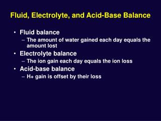
Introduction • Fluid & electrolytes are fundamental biochemical systems • Tightly controlled homeostatic mechanisms • Simple and cheap analytical processes • Underlying physiology complex
Water distribution Intracellular H2O (28L) Extracellular H2O (14L) Plasma Potassium (4 mmol/L) • Total adult water content – 42L • 60% body weight (men) • 55% body weight (women) Potassium (110 mmol/L) Sodium (135 mmol/L) Sodium (10mmol/l 3.5L Na+,K+,ATPase
Water balance Water OUT (obligatory) • Skin 500ml • Lungs 400ml • Gut 100ml • Kidney 500 ml Water IN • Metabolism 400mL • Diet 1100mL Total in 1500mL = Total out 1500mL
Control of water balance • Thirst • Fluid shifts between ICF and ECF • Anti Diuretic Hormone (ADH) or vasopressin In response to changes in: ECF Osmolarity (sensed by osmoreceptors) Osmolarity – measure of solute concentration (no. of moles of solute per unit volume of solution (Osm/L))
Action of ADH • Released from posterior pituitary • Acts on renal collecting ducts to allow • re-absorption of water • Primary aim is to keep ECF osmolarity constant • BUT volume depletion – ECF volume maintained at expense of osmolarity
Water homeostasis Water depletion
Water homeostasis Normal Water overloaded Serum osmo <290mosm/L Urine osmo <100mosm/L Serum osmo = 290mosm/L Urine osmo = 100-600mosm/L Dehydrated Serum osmo >290mosm/L Urine osmo >600mosm/L
Sodium balance Sodium OUT (Obligatory losses) • Gut/skin 10 mmol (Loss dependent on intake) • Kidney 90–190mmol Sodium IN • Diet 100-200 mmol
Control of sodium balance • Renin – angiotensin – aldosterone system
Aldosterone • Produced by adrenal • Acts on renal distal tubule to increase re-absorption of sodium (in exchange for K+ / H+) In response to changes in: ECF Volume (sensed by baroreceptors)
Sodium content vs concentration • ECF Na content determines ECF volume • Na content leads to hypervolaemia • Na content leads to hypovolaemia • [Na+] reflects water balance NOT sodium balance (in most cases) • [Na+] = water depletion (dehydration) • [Na+] = water overload • Na content may be normal, low or high
Electrolyte Disturbances • Hypernatraemia • Inadequate fluid intake • Diabetes insipidus • Pituitary - ADH deficiency • Nephrogenic – ADH resistance • Hyponatraemia • Excessive fluid intake / administration • Impaired water excretion ( ADH) • Physiological - response to hypovolaemia • Pathological - SIADH (Syndrome of Inappropriate ADH Secretion)
Hyponatraemia • Sodium deplete (hypovolaemic) (2º ADH and H2O overload) • Mineralcorticoid deficiency, e.g., adrenal insufficiency • Diarrhoea / vomiting • Diuretics • Na-losing nephropathy • Sodium overload (hypervolaemic) (2º ADH and H2O overload) • Cirrhosis • Renal failure • Heart failure • Nephrotic syndrome • Normal sodium balance (normovolaemic) • Cortisol deficiency, hypothyroidism, renal failure • SIADH – drugs, tumours, chest infections, CNS (excessive ADH secretion)
Potassium balance • Potassium OUT • (Obligatory losses) • Faeces 5-10 mmol • Skin 5-10 mmol • (Loss dependent on intake) • kidney 40-190 mmol Potassium IN • Diet 60-200 mmol Kidney – main regulator of total body potassium Aldosterone allows excretion of K+ in exchange for Na+
Potassium distribution • Intra-cellular cation • Plasma [K+] poor indicator of total body K+ • Potassium moves in and out of cells due to: • Hormonal control, e.g., insulin • Reciprocal movement of H+
Electrolyte Disturbances Hypokalaemia • Low intake – oral (rare), parenteral • K+ into cells • Insulin, theophylline, catecholamines • Alkalosis • Increased losses • Gut – diarrhoea, laxative abuse, vomiting • Kidneys – Mineralocorticoid excess, renal tubular defects
Electrolyte Disturbances Hyperkalaemia • Increased intake (+ impaired excretion) • Out of cells • Insulin deficiency • Acidosis • Cell breakdown – rhabdomyolysis, tumour lysis • Impaired excretion • Renal failure • Mineralocorticoid deficiency • Drugs - ACEi, K+ sparing diuretics • Acidosis
Analytical parameters • Serum / plasma • Na • K • Osmolarity (osmolar gap) • Urine • Na • K • Osmolarity
Osmolarity • Osmolarity (osm/L) vs osmolality (osm/Kg) • Osmolality is measured (NOT temperature dependent) • If concentration of solutes is low: osmolality osmolarity • Calculated osmo =2[Na+]+[K+]+[urea]+[gluc] • Osmolar gap = Measured osmo – calculated osmo • Normal range 10 – 15 mmol / L • Increased osmolar gap due to e.g., ethanol, methanol, ethylene glycol
Indications for measurement (1) • Serum Na / K • Renal function • Fluid status • Adrenal function • Pituitary function • Drug side effects • Acute illness (e.g., DKA, severe V&D) • Nutritional status (e.g., TPN) • Urine Na / K • Investigation of hyponatraemia / hypokalaemia • TPN
Indications for measurement (2) • Serum Osmo • Verification of true hyponatraemia • Investigation of diabetes insipidus* • ?Poisoning / alcohol • Urine Osmo • Investigation of hyponatraemia • Investigation of diabetes insipidus* *May be as part of water deprivation test
Water Deprivation Test (1) • Investigation of Diabetes Insipidus (DI) • Principle: Deprive patient of fluids to allow serum osmo to rise and see whether urine concentrates (i.e., urine osmo increases). • Protocol: • Patient usually fasted overnight. May or may not be allowed fluids overnight. • Serum and urine osmo measurements performed approx every hour (and patient’s weight and urine volume recorded)
Water Deprivation Test (2) • End points: serum osmo > 300 mosm/L or >5 % loss of body weight • Urine osmo > 600 mosm/L DI excluded • Urine osmo < 200 mosm/L DI diagnosed • Urine osmo 200-600 equivocal • If DI diagnosed, synthetic ADH (DDAVP) given nasally. • Urine osmo > 600 mosm/L pituitary DI • Urine osmo < 200 mosm/L nephrogenic DI
Ion selective electrodes Na+ Na+ K+ Na+ K+ • Ion selective membrane • Na+ (glass), K+ (valinomycin) • Ions interact with electrode to create potential difference • Produces a current, which is proportional to [Na+]
Direct vs indirect ISE • Direct ISE (e.g., Li analyser) • Measures activity of Na+ in neat sample • Unaffected by electrolyte exclusion effect • Unsuitable for urine analysis • Indirect ISE (e.g., Roche Modular) • Measures activity of sample diluted in high ionic strength buffer • Suitable for urine analysis • Unsuitable for whole blood • Affected by electrolyte exclusion effect
Electrolyte exclusion effect • Normal serum contains 93 % water • Water content lower in lipaemic or high protein concentration samples • Spuriously low [Na+] in e.g., lipaemic samples when analysed using indirect ISE Treat sample with lipoclear, then analyse using direct ISE
Osmometry • Freezing point depression principle • The freezing point of a solvent lowers when a solute is added to aqueous solutions • One osmole of solute per Kg of solvent depresses the freezing point by 1.85 °C
Artefactual hyponatraemia • Electrolyte exclusion effect (indirect ISE) • Lipaemic samples or high total protein • Normal serum osmo • Measure on direct ISE • Hyperosmolar hyponatraemia • Very high glucose (high serum osmo) • Causes fluid shifts from ICF to ECF, which dilutes [Na+] • Artefactual – does not require treatment
Artefactual hyperkalaemia • Causes • Haemolysed • On cells (worse at 4ºC) • EDTA contamination • Very high WCC or platelets • Integrity checks • Haemolysis index • Sample date / time • Calcium / Mg • Check FBC, repeat in LiHep if necessary
Reference ranges • Na 133 – 146 mmol/L • K 3.5 – 5.3 mmol/L
Panic ranges (1) • Na+ >155 mmol/L • Thirst, difficulty swallowing, weakness, confusion • Na+ <120 mmol/L • Weakness, postural dizziness, behavioural disturbances, confusion, headache, convulsions, coma Rate of change of [Na+] important
Panic ranges (2) • K+ >6.5 mmol/L • Increased risk of sudden cardiac death • K+ <2.5 mmol/L • Weakness, constipation, depression, confusion, arrhythmias, polyuria
Case example - 1 • 48 y female • Partial ptosis (drooping of eyelid) • Na 144 mmol/L (133 – 146) • K +7.0 mmol/L (3.5 – 5.3) • Urea 4.5 mmol/L (2.5 – 7.8) • Creat 65 µmol/L (44 – 133) • eGFR 85 mL/min (>90)
Case example - 1 • Check sample • ?Haemolysed – NO • Date/time – OK • Ca/Mg added • Ca -1.0 mmol/L (2.2–2.6) • Mg -0.11 mmol/L (0.7 – 1.0) • EDTA contamination
Case example - 2 • 17 y female • 2 month hx lethargy and tiredness • Dizzy on standing • Pigmentation in mouth and in palmar creases • BP 120/80 mmHg lying, fell to 90/50 mmHg when standing
Case example - 2 • Na -128 mmol/L (133-146) • K +5.4 mmol/L (3.5-5.3) • Urea +8.5 mmol/L (2.5-7.8) • Creat 55 µmol/L (44-133) • Fasting glucose -2.5 mmol/L
Case example - 2 • Short Synacthen test • 09:00 h 150 nmol/L • 09:30 h 160 nmol/L • 10:00 h 160 nmol/L (Normal response: cortisol >550 nmol/L, with increase of >200 nmol/L) • ACTH 500 ng/L (<50) • High titre anti-adrenal antibodies
Case example - 2 • Primary adrenal insufficiency Hypothalamus CRH CRH Pituitary ACTH ACTH Adrenal Cortisol Cortisol
Case example - 2 • Addison’s disease (autoimmune adrenal insufficiency) • Led to hyponatraemia • Lack of aldosterone – uncontrolled Na loss from kidneys • Hypovolaemic - 2° increase in ADH and water retention • Treatment: mineralocorticoid (aldosterone) and glucocorticoid (cortisol) rx