Download
1 / 45
780 likes | 2.04k Vues
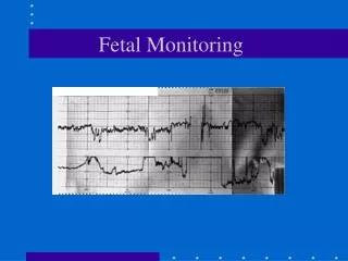
Fetal Monitoring Basics Expanded. NUR 231 M. Johnston, RN-BC, M.Ed. Cindy Irwin , RNC, MN. Types of Monitoring. Auscultation- listen to fetal heart rate (FHR) Electronic Fetal Monitoring – use of instruments to record FHR and uterine contractions(U/Cs). Auscultation.

E N D
Fetal Monitoring Basics Expanded NUR 231 M. Johnston, RN-BC, M.Ed. Cindy Irwin , RNC, MN
Types of Monitoring Auscultation- listen to fetal heart rate (FHR) Electronic Fetal Monitoring – use of instruments to record FHR and uterine contractions(U/Cs)
Auscultation • Doppler - ultrasound converts sounds waves to signals of fetal heart • Fetoscope- Like stethoscope, open end pressed on abdomen, used less frequently
Electronic Fetal Monitoring • Measures response of FHR to uterine contractions (U/Cs) • Intermittent or Continuous • External • Ultrasound transducer • Tocotransducer • Internal • Fetal Scalp Electrode • Intrauterine Pressure Catheter
Fetal Heart Rate Characteristics • Evaluate to determine fetal status • NICHD terminology • Baseline Rate • BaselineVariability • Accelerations (present or absent) • Decelerations (present or absent) • Changes or trends over time
Baseline (BL) • Normal range 110-160 bpm • Measure between U/Cs for at least 2 min. period during 10 minute segment • Tachycardia - >160 bpm for >10 minutes • Bradycardia - <110 bpm for >10 minutes
Classifications of FHR Variability • Fluctuations in FHR, irregular in frequency and amplitude • Absent 0-2 bpm • Minimal >2 <6 bpm • Moderate 6 -25 bpm • Marked >25 bpm
Periodic/Episodic ChangesPeriodic with uterine contractionsEpisodic without uterine contractions
Accelerations • Abrupt increase in FHR above BL • Present or Absent • < 32 wks gestation • Peak ≥ 10 bpm above BL for at least 10 sec. • >32 wks gestation • Peak ≥ 15 bpm above BL for at least 15 sec. • Accel ≥ 10 min. is defined as BL change
Accelerations • Abrupt increase in FHR above BL • Peak ≥ 15 bpm above BL for at least 15 sec.
Recognition Criteria for Fetal Heart Rate Accelerations • Transient increase in the Fetal Heart Rate • > 32 weeks acceleration stays 15 beats above the baseline for at least 15 seconds • < 32 weeks acceleration stays 10 beats above the baseline for at least 10 seconds
Types of Decelerations • Early – Gradual decrease and return to BL,mirrors the U/C • Variable – Abrupt (<30 sec) decrease (≥15 sec down, lasting ≥ 15 sec and <2 min from onset to return to BL) • Late – Gradual decrease (≥30 sec) and gradual return to BL; delayed timing nadir occurs after peak of U/C • Prolonged – Decrease in FHR below BL ≥15 sec, lasting ≥ 2 min. but <10 min.
Early Deceleration • Gradual decrease and return to BL • Mirrors the U/C
Early Decelerations • Usually benign • May be associated with descent of fetus • Monitor to assure fetal well-being, no evidence of worsening condition
Variable Deceleration • Abrupt (<30 sec) decrease (≥ 15 sec down, lasting ≥ 15 sec and < 2 min. from onset to return to BL)
Variable Deceleration- Cause and Treatment Cause- • Umbilical cord compression resulting in baroreceptor stimulation Treatment- • Assess baseline variability, rate • Reposition mother • Notify provider • Check for cord prolapse • Apply internal monitors • or turn off oxytocin • Administer O2 by mask • Prepare for possible amnioinfusion • Document interventions/FHR response
Late Deceleration • Gradual decrease (≥ 30 sec) and gradual return to BL • Delayed timing, nadir occurs after peak of U/C
Late decelerations - Cause and Treatment Cause- Placental insufficiency Treatment • Assess baseline variability, rate, accelerations • Reposition mother on side • IV fluids • or turn off oxytocin • Notify provider • Administer O2 by mask • Apply internal monitors • Evaluate scalp stimulation • Document interventions/FHR response • Exit plan
Prolonged Deceleration • Decrease in FHR below BL ≥ 15 sec, lasting ≥ 2 min. but < 10 min.
Causes of Prolonged Decelerations • Uterine hyperstimulation or hypertonus • Abruptio placenta • Acute maternal hypotension • Uterine rupture • Maternal hypoxia • Umbilical cord accidents • Terminal fetal conditions • Vasaprevia • Rapid fetal descent • Vagal stimulation or maternal Valsalva
Treatment for Prolonged Decelerations • Notify provider • Assess baseline variability, rate, accelerations Reposition mother on side • IV fluids • or turn off oxytocin • Administer O2 by mask • Apply internal monitors • Do not attempt scalp stimulation • Document interventions/FHR response • Exit plan
Fetal Heart Rate Patterns Indeterminate FHR tracings that do not meet the criteria for Normal or Abnormal • Abnormal • Absent baseline variability • and any of the following: • Recurrent late decels • Recurrent variable decels • Bradycardia • or • Sinusodial pattern • Normal • ALL required: • Moderate variability • Baseline rate 110-160 • No late or variable decels • Early decels present or • absent • Accels: present or absent Category I Strongly associated with normal acid base status Category II Not predictive of abnormal fetal acid base status but inadequate evidence to classify as normal or abnormal Category III Predictive of abnormal fetal acid base status
Fetal Heart Rate Interpretation System Normal Indeterminate Abnormal
FHR Interpretation • Information about fetal oxygenation/placental function • Somewhat subjective • Abnormal patterns may need further testing
Monitoring Uterine Contractions • Assess U/C pattern while assessing FHTs • External • Palpation • EFM Toco measures frequency, duration • Noninvasive • Internal • Intrauterine pressure catheter (IUPC) • Measures exact intrauterine pressure • Invasive
Why Monitor? • FHR changes in response to oxygenation, gestation, and certain stimuli • EFM provides more objective data than auscultation • Infers information about current and ongoing fetal oxygenation
Interventions • Abnormal FHR pattern: • Notify provider • Change maternal position • Give oxygen via mask • Increase IV fluids • Consider medication to relax uterus
Other Fetal Surveillance • Non-Stress Test (NST) - EFM • Contraction Stress Test (CST) - EFM • Biophysical Profile (BPP) - U/S • Doppler Flow Studies/Growth - U/S • Fetal Movement Count-maternal sensation/palpation
Intermittent Auscultation • What’s the evidence? • ACOG, AWHONN support the use of auscultation as an appropriate way to evaluate fetal heart rate for the uncomplicated patient • Neonatal outcomes comparable to those with use of EFM based on randomized clinical trials
Technique for IA • Assess contractions by palpation • Determine fetal position • Determine maternal pulse rate • Place Doppler over fetal back or thorax • Determine baseline FHR by listening between contractions for 30-60 seconds: differentiate from maternal HR • Count FHR immediately after contraction for 30-60 seconds • Chart under Intermittent Auscultation- Baseline, Rhythm, Increases, Decreases
How often? On admission, obtain 20 minute FM tracing If Category I tracing, no risk factors present, and provider order for IA: Document FHR and uterine activity: • Latent phase: As ordered • Active labor: Every 15-30 minutes • Second stage: Every 5-15 minutes
Comparison Model for Palpation of Uterine Activity PALPATION OF UTERUS FEELS LIKE CONTRACTION INTENSITY • Easily indented • Tip of nose • Mild • Can slightly indent • Chin • Moderate • Cannot indent • Forehead • Strong
Limitations • Difficult to hear FHR with if pt obese, has an increased AFI, or with maternal or fetal movement • No tracing to review at a later time • Certain EFM characteristics cannot be measured (sinusoidal pattern) • Requires practice
Benefits • Lower C/S and operative delivery rates compared to EFM for patients without risk factors • Allows maternal freedom of movement/ambulation • Increased hands-on contact with patient • Increased patient satisfaction