Download
1 / 72
750 likes | 1.32k Vues
BASIC PATHOLOGICAL ASPECTS OF NERVOUS SYSTEM PATHOLOGY. Esti D. S. Soetrisno B. Rino Pattiata Departement Anatomic Pathology Faculty of Medicine University of Indonesia. BASIC PATHOLOGICAL MANIFESTATION OF SOME DISTURBANCES. DYS – NEURO EMBRYOGENESIS

E N D
BASIC PATHOLOGICAL ASPECTS OF NERVOUS SYSTEM PATHOLOGY Esti D. S. Soetrisno B. Rino Pattiata Departement Anatomic Pathology Faculty of Medicine University of Indonesia
BASIC PATHOLOGICAL MANIFESTATION OF SOME DISTURBANCES DYS – NEURO EMBRYOGENESIS • ABORTION / INTRA-UTERINE FETAL DEATH (IUFD) • ABNORMALITIES : TERATOGENIC, MONSTER, CONGENITAL ANOMALY AGENESIS : There is no processus (anlage) of all or partial part of NS No formation of NS IUFD APLASIA : There is only NS Streak Formation abortion HYPOPLASIA : Failure to growth of all or partial part of NS Hypotrophy (Micro Insize) Hypofunction / Fatal e.g Microensephaly, Arnold – Chiary Syndrome
Hyperplasia : Overgrowth parts of NS e.g Macroensephaly, Hydrocephallus, Function? Hypertrophy: True Hypertrophy / Pseudo Hypertrophy • Defect On Enclosing of the Neural Tube There is “Cele” Formation, or Spina Bifida Formation (Occulta/Aperta) e.g Meningocele, Encephalo -/ Myelo – Meningcocele, Syringo -Encephalo –/ Myelo – Meningcocele (Syringo Myelia)
DYS – HISTOGENESIS : incorrect migration and/or naturation – differentation ECTOPIC : mature tissue found in abnormal places HETEROPIC : intermingled of some mature tissuesin abnormal places HAMARTOMA : abnormal composition of mature tissues at its normal places NEOPLASMA (GEN MUTATION) : benign and malignant
DYS – NEUROANATOMY Abnormalities of anatomy / location of NS - Dyslocation - Reverse of Several Centre DYS - NEUROCHEMISTRY - NEUROPHYSIOLOGY INHIBIT : Slow Conduction – Slow Movement / Analysis / etc EXCITE : Rapid / Hyperactivity (ies) DYS – REGULATION / CONTROL : UNCONTROL MOVEMENT – PATHOLOGICAL REFLEXES DYSFUNCTIONAL IMPULS CONDUCT
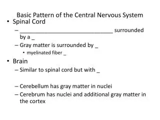
CNS CELLS • NEURON • GLIAL CELL • ASTROCYTE • OLIGODENDROGLIA • EPENDYMA • MICROGLIA • CHOROID PLEXUS CELL
OLIGO DENDROGLIA NEURON ASTROCYTE
Neuron • Effector cells of Nervous System • Neuron loss with progressive aginh • Neuron of CNS cannot effectively regenerate axons over long distance → limit ability of CNS to respond to different type of injury • Infarct transects internal capsule creates permanent motor deficiti • Neuron in CNS don’t remyelinate → demyelinating disease causes permanent functional deficit (multipel sclerosis)
PIGMENTED NEURON ( SUBSTANTIA NIGRA ) neuromelanin
ATROPHIC NEURON hyperchromatic Loss of neurons * global/regional reduction (atrophic) * single neuron
ATROPHIC CEREBRAL CORTEX
CHROMATOLYSIS Injured neuron swell → cytoplasm swell → chromatolysis: response to injury Reversible/death CYTOPLASM FLUID ACCUMULATION MARGINATION NUCLEUS NISSL SUBSTANCE
CENTRAL CHROMATOLYSIS ANTERETROGADE DEGENERATION
Astrocyte • Support neurons • Promote repair
GLIOSIS • Reaction to injury • Proliferation of astrocyte • Evolves in hours to day and persists to an extent that is usually commensurate with the severity of injury • Reactive astrocyte : gemistocytic astrocyte: exentric plump nuclei, eosinophilic cytoplam • Glial scar: composed of reactive astrocytes and their processes.
OLIGODENDROGLIA • Neuroectodermal origin • Myelin-producing cells during late gestational period and early neonatal
EPENDYMA • Modulate fluid transfer between the cerebrospinal fluid and CNS • During gestation some viral target the ependymal cell → aqueductus stenosis → congenital hydrocephalus
CANALIS CENTRALIS EPENDYM
MICROGLIA • Phagocytic macrophage-derived cells • Reactions: changes in areas of injury • 2 pattern : focal and diffuse microgliosis • Microglial nodule: responses to viral or other infection. • Rod cells: prominent elongated nucleus • Gitter cells: response to necrosis: it will become phagocytic, accumulate lipid and other material
ACTIVATED MICROGLIA MYELINOLYSIS
INTRA NUCLEAR INCLUSION ( CYTOMEGALO VIRUS )
NEGRI BODY INTRACYTOPLASM (RED) (RABIES ENCEPHALITIS)
VASCULAR DILATATION (HYPEREMIA) PMN NEUTROPHIL NEURONOPHAGIA
HYDROCHEPALUS • TYPE : • COMMUNICANS : obstruction occurs outside ventricle system • NON-COMMUNICANS • EXVACUO (COMPENSATED)
HYDROCEPHALUS • Primary hydrocephalus • Accompanied by increased intracranial pressure • Due to: • Obstruction • Congenital • acquired • Impaired CSF absorption • Excess CSF production • Secondary hydrocephalus • Compensatory to loss of cerebral tissue
SITES OF OBSTRUCTION OF CSF PATHWAY • Subarachnoid space • Arachnoid granulationes • Plexus choroid • Lateral ventricle • 3rd ventricle • Cerebral aqueduct • 4th ventricle • Exit foramina
OBSTRUCTED AQUADUCT SYLVIOUS ( BRAIN TUMOR)
OBSTRUCTIVE HYDROCEPHALUS ( NEOPLASM )
OBSTRUCTIVE HYDROCEPHALUS ( INFECTION )
OBSTRUCTIVE HYDROCEPHALUS ( GLIAL TISSUE POST VIRAL INFECTION)
TRAUMA • Penetrating wounds produce hemorrhage and blast effects. Velocity contributes a blast effect to a projectile • High-velocity : it disrupts tissues by its own mass and also centrifugal blast that enlarges the diameter → immediate death • Low-velocity • Seizures are threat in healed penetrating wounds, 6-12 mo after : collagenous tissue is displaced in the brain
HEMORRHAGIC TRACT (PENETRATING WOUND)
Subdural hematoma • Significant cause of death from falls, assaults, vehicular acidents, sporting mishaps • Frontal/occipital area is struck by blunt object → cerebral hemispher displaced in an anteroposterior direction → hit against inner aspect • Soft cerebral tissue becomes compact then recoil → shearing effect • Usually stop after 25-30 mL
Subdural hematoma • Tissue response • Formation of granulation tissue → outer membrane • Fibroblast from outer membrane moved into the hematoma → inner membrane : 2 weeks • Evolution: • Reabsorbe leave a small amount of telltale hemosiderophage • Remain static, with potential for calcification • Enlarge : 6 months
CHRONIC SUBDURAL HEMATOMA (INNER NEOMEMBRANE)
EPIDURAL HEMATOMA • Middle meningeal artery branches splay across temporal-parietal area • Hemorrhage into epidural space, separating dura from calvaria • 4-8 hours: asymptomatic • 30-50 mL: intracranial pressure increased → exceed venous pressure → circulatory stagnation and cerebral ischemia → global cerebral hypoxia
EPIDURAL HEMATOMA • Cushing reflex : protective response • HR slow to increase ventricular filling • Myocardial contraction is forceful • Systolic pressure increased • Compensatory mechanism exhausted : temporal lobe displaced downward → transtentorial herniation • Herniation compress uncus/hyppocampus against midbrain and other structures : 3rd cranial nerve • Pupil fixed and dilated
EPIDURAL HEMATOMA (FRONTO PARIETAL)
HERNIATION • Cingulate gyrus under falx cerebri • Hippocampal uncus and parahippocampal gyrus over tentorium cerebeli • Cerebelar tonsilar through foramen magnum • Any defect in the dura and skull SITES OF HERNIATION
TRANSTENTORIAL HERNIATION (MIDBRAIN DISPLACED)