Download
1 / 86
860 likes | 1.17k Vues
ORAL CAVITY-1. This resource is licensed under the Creative Commons Attribution Non-Commercial & No Derivative Works License. Objectives. Recognise and describe a section of the lip , identifying the main features and constituent tissues.

E N D
ORAL CAVITY-1 This resource is licensed under the Creative Commons Attribution Non-Commercial & No Derivative Works License
Objectives Recognise and describe a section of the lip, identifying the main features and constituent tissues. Recognise and describe a section of the palate, distinguishing between hard and soft palate and identifying its epithelia and glands. Recognise and describe a section of tongue (low power) appreciating the arrangement of epithelium, connective tissue (with fat cells), blood vessels, nerves, glands and muscle. Recognise and describe the main lingual papillae (high power). To be able to distinguish and describe serous and mucous lingual glands.
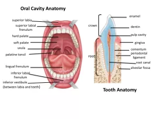
SLIDE 1 Lip (cat) At low magnification identify: oral surface aboral surface labial margin 1.0 mm
SLIDE 1 Lip (cat) oral surface labial margin What type of epithelium covers : 1. the oral surface? 2. the aboral surface? 1.0 mm aboral surface
What type of epithelium covers : 1. the oral surface? stratified squamous (little keratinisation) 2. the aboral surface? SLIDE 1 Lip (cat) oral surface labial margin 1.0 mm aboral surface
What type of epithelium covers : 1. the oral surface? stratified squamous (little keratinisation) 2. the aboral surface? keratinised stratified squamous SLIDE 1 Lip (cat) oral surface labial margin 1.0 mm aboral surface
SLIDE 1 Lip (cat) At low magnification identify; on the aboral surface: hair follicles of two types; what are they? 1.0 mm
SLIDE 1 Lip (cat) At low magnification identify; on the aboral surface: hair follicles of two types; what are they? Sinus hair follicles and compound hair follicles. sinus hair follicles 1.0 mm compound hairs
SLIDE 1 Lip (cat) 100 µm Identify: two types of gland. What are they? 1.0 mm
B SLIDE 1 Lip (cat) A 100 µm Identify: two types of gland. What are they? A : sebaceous glands. B : apocrine sweat glands. B A 1.0 mm
SLIDE 1 Lip (cat) What cranial nerve might be seen in this section? 1.0 mm
SLIDE 1 Lip (cat) What cranial nerve might be seen in this section? The facial or seventh cranial nerve. 150 µm 1.0 mm
SLIDE 1 Lip (cat) What skeletal muscle is probably within this section? 1.0 mm
SLIDE 1 Lip (cat) muscle connective tissue What skeletal muscle is probably within this section? The orbicularis oris muscle. 1.0 mm
SLIDE 1 Lip (cat) How are these muscle fibres orientated re. the plane of the section? 50 µm 1.0 mm
SLIDE 1 Lip (cat) How are these muscle fibres orientated re. the plane of the section? In transverse section. 50 µm 1.0 mm
SLIDE 1 Lip (cat) What is this muscle’s function? 50 µm 1.0 mm
SLIDE 1 Lip (cat) What is this muscle’s function? It closes the lips. 50 µm 1.0 mm
SLIDE 2 Palate (mouse) This is a sagittal section through the head of a very young mouse. Identify: brain in cranium nasal cavity ethmoid bone (cribriform plate) hard palate soft palate skin nasopharynx palatine bone 1.0 mm
SLIDE 2 Palate (mouse) ethmoid bone nasal turbinates skin brain This is a sagittal section through the head of a very young mouse. Identify: brain in cranium nasal cavity ethmoid bone (cribriform plate) hard palate soft palate skin nasopharynx palatine bone nasal cavity palatine bone hard palate nasopharynx 1.0 mm soft palate
SLIDE 2 Palate (mouse) Another section through the head. In this section only the hard palate is present. Identify: nasal cavity ethmoid bone (cribriform plate) hard palate skin palatine bone incisor 1.0 mm
SLIDE 2 Palate (mouse) ethmoid bone nasal turbinates nasal cavity skin with hairs Another section through the head. In this section only the hard palate is present. Identify: nasal cavity ethmoid bone (cribriform plate) hard palate skin palatine bone incisor nasal opening incisor tooth palatine bone 1.0 mm hard palate
Sagittal section of horse head. With reference to the dissection class : locate the areas of hard and soft palate.
Oral Cavity _ Dog ▪ Tongue & larynx have been removed ▪ Soft palate has been incised along the palatoglossal arches ▪ Note partially pigmented mucosa of upper lip ▪ Note ridged, pigmented mucosa of hard palate ▪ Identify : soft palate, junction of hard and soft palates SLIDE 2 Palate (mouse) What type of epithelium covers the hard palate? 250 µm
SLIDE 2 Palate (mouse) Stratum : 1: basale 2: spinosum 3: granulosum 4: lucidum (unclear) 5: corneum keratinised 1 What type of epithelium covers the hard palate? Keratinised stratified squamous epithelium. 2 3 4 5 100 µm
SLIDE 2 Palate (mouse) nasopharynx What type of epithelium covers the oral surface of the soft palate? oral cavity 250 µm
SLIDE 2 Palate (mouse) What type of epithelium covers the oral surface of the soft palate? Stratified squamous epithelium. stratified squamous epithelium, some keratinisation oral cavity 50 µm
SLIDE 2 Palate (mouse) 250 µm Junction between soft and hard palate. What glands are present in the soft palate? 50 µm
SLIDE 2 Palate (mouse) 250 µm Junction between soft and hard palate. What glands are present in the soft palate? Palatine glands (minor salivary gland). palatine glands 50 µm
SLIDE 2 Palate (mouse) aboral surface What type of epithelium covers the aboral surface of the soft palate seen here? 25 µm
SLIDE 2 Palate (mouse) ciliated, pseudostratified, columnar epithelium aboral surface What type of epithelium covers the aboral surface of the soft palate seen here? Ciliated, pseudostratified columnar epithelium. 25 µm
SLIDE 2 Palate (mouse) ciliated, pseudostratified, columnar epithelium aboral surface What type of epithelium covers the aboral surface of the soft palate seen here? Ciliated, pseudostratified columnar epithelium. What striated muscle might be sectioned in the soft palate? 25 µm
SLIDE 2 Palate (mouse) ciliated, pseudostratified, columnar epithelium aboral surface What type of epithelium covers the aboral surface of the soft palate seen here? Ciliated, pseudostratified columnar epithelium. What striated muscle might be sectioned in the soft palate? Palatine muscle. striated muscle palatine gland 25 µm
Embryonic SLIDE Demo TS Head SECTION THROUGH ORAL AND NASAL CAVITIES Identify: 1. Oral cavity. 2. Nasal cavity. 3. Nasal septum. 4. Nasal conchae. 5. Tongue. 6. Mandible. 7. Developing molars. 1.0 mm
Embryonic SLIDE Demo TS Head SECTION THROUGH ORAL AND NASAL CAVITIES 4 2 2 3 Identify: 1. Oral cavity. 2. Nasal cavity. 3. Nasal septum. 4. Nasal conchae. 5. Tongue. 6. Mandible. 7. Developing molars. 1 7 5 6 1.0 mm
SLIDE 3 Tongue (cat) Whole section seen at low magnification. Identify: epithelium fat (adipose) tissue large blood vessels and nerves glands lingual frenulum striated muscle- seen in longitudinal, oblique and transverse orientations. 5.0 mm
Tongue, larynx & pharynx – dog ▪ Dorsal view ▪ Pharynx and proximal oesophagus have been opened dorsally. Window has been cut in trachea. ▪ Identify : papillated (filiform) mucosa of tongue root, epiglottis, arytenoid cartilages & musculature, vestibulum oesophagi*, piriform recess*, limen pharyngoesophageum*, aditus laryngis, thyrohyoid bones, stylohyoid bones (transected). * Pars laryngea pharyngis.
Oral Cavity - Sheep 2 ▪ Skin reflected from mandible, partially reflected from maxilla. M. buccinator removed. left mandible in situ, right mandible has been transected just caudal to the mandibular symphysis; and removed following reflection of masseter, temporal & pterygoid mm. (latter apparent dorsal to the tongue) from the mandibular ramus. 1 2 1 ▪ M. buccinator has been transected and reflected to expose gingival mucosa, buccal mucosa, tongue, premolar & molar teeth. ▪ Identify papillae of various sorts of the lingual and buccal mucosa. ▪ Tongue reflected ventrally to show papillated mucosa (what sort of papillae are apparent?) & torus linguae. ▪ Note : pigmented, ridged mucosa of hard palate, non-pigmented soft palate, mandibular incisor teeth, absence of upper incisors.
5.0 mm SLIDE 3 Tongue (cat) dorsal Whole section seen at low magnification. Identify: epithelium lateral
SLIDE 3 Tongue (cat) lateral surface dorsal surface What type of epithelium covers the tongue? 100 µm
SLIDE 3 Tongue (cat) lateral surface dorsal surface What type of epithelium covers the tongue? Keratinised stratified squamous epithelium. 100 µm
SLIDE 3 Tongue (cat) lateral surface dorsal surface On which surface of the tongue is the epithelium most keratinised and why? 100 µm
SLIDE 3 Tongue (cat) lateral surface dorsal surface On which surface of the tongue is the epithelium most keratinised and why? On the dorsal surface. There is most contact on this surface. 100 µm
SLIDE 3 Tongue (cat) Whole section seen at low magnification. Identify: glands 5.0 mm
SLIDE 3 Tongue (cat) Some minor salivary glands (lingual) can be seen in this section. salivary gland 0.5 mm
SLIDE 3 Tongue (cat) duct Minor salivary glands in body of the tongue. See also slide 4. acini 50 µm
SLIDE 3 Tongue (cat) Whole section seen at low magnification. Identify: fat (adipose) tissue striated muscle- seen in longitudinal, oblique and transverse orientations. lingual frenulum frenulum 5.0 mm
SLIDE 3 Tongue (cat) At fairly low magnification, the bundles of striated fibres forming the intrinsic muscles of the tongue can be seen cut in many orientations. 250 µm
SLIDE 3 Tongue (cat) adipocytes Here at medium magnification striated muscle fibres can be seen cut in transverse and longitudinal section. Note the large areas of unilocular adipocytes (white fat). striated muscle A : TS section B : LS section B A 100 µm
SLIDE 3 Tongue (cat) Whole section seen at low magnification. Identify: large blood vessels and nerves. 5.0 mm