Download
1 / 1
10 likes | 141 Vues
Crystallography with a dual source diffractometer at Reading

E N D
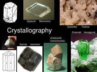
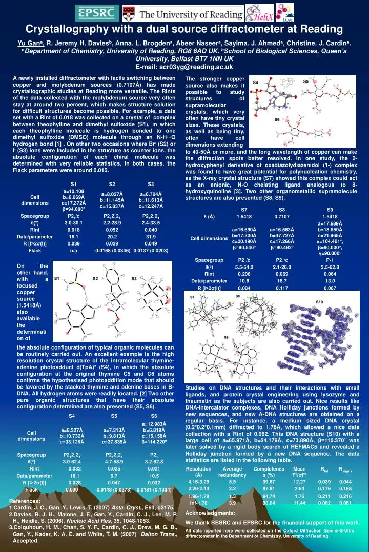
Crystallography with a dual source diffractometer at Reading Yu Gana, R. Jeremy H. Daviesb, Anna. L. Brogdena, Abeer Naseera, Sayima. J. Ahmeda, Christine. J. Cardina. aDepartment of Chemistry, University of Reading, RG6 6AD UK. bSchool of Biological Sciences, Queen’s University, Belfast BT7 1NN UK E-mail: scr03yg@reading.ac.uk A newly installed diffractometer with facile switching between copper and molybdenum sources (0.7107Å) has made crystallographic studies at Reading more versatile. The Rints of the data collected with the molybdenum source very often stay at around two percent, which makes structure solution for difficult structures become possible. For example, a data set with a Rint of 0.018 was collected on a crystal of complex between theophylline and dimethyl sulfoxide (S1), in which each theophylline molecule is hydrogen bonded to one dimethyl sulfoxide (DMSO) molecule through an N-H∙∙∙O hydrogen bond [1] . On other two occasions where Br- (S2) or I- (S3) ions were included in the structure as counter ions, the absolute configuration of each chiral molecule was determined with very reliable statistics, in both cases, the Flack parameters were around 0.015. The stronger copper source also makes it possible to study structures of supramolecular crystals, which very often have tiny crystal sizes. These crystals, as well as being tiny, often have cell dimensions extending to 40-50Å or more, and the long wavelength of copper can make the diffraction spots better resolved. In one study, the 2-hydroxyphenyl derivative of oxadiazolydiazenidol (1-) complex was found to have great potential for polynucleation chemistry, as the X-ray crystal structure (S7) showed this complex could act as an anionic, N-O chelating ligand analogous to 8-hydroxyquinoline [3]. Two other organometallic supramolecule structures are also presented (S8, S9). On the other hand, with a focused copper source (1.5418Å) also available, the determination of the absolute configuration of typical organic molecules can be routinely carried out. An excellent example is the high resolution crystal structure of the intramolecular thymine-adenine photoadduct d(TpA)* (S4), in which the absolute configuration at the original thymine C5 and C6 atoms confirms the hypothesised photoaddition mode that should be favored by the stacked thymine and adenine bases in B-DNA. All hydrogen atoms were readily located. [2] Two other pure organic structures that have their absolute configuration determined are also presented (S5, S6). Studies on DNA structures and their interactions with small ligands, and protein crystal engineering using lysozyme and thaumatin as the subjects are also carried out. Nice results like DNA-intercalator complexes, DNA Holliday junctions formed by new sequences, and new A-DNA structures are obtained on a regular basis. For instance, a medium sized DNA crystal (0.2*0.2*0.1mm) diffracted to 1.78Å, which allowed a nice data collection with a Rint of 0.062. This DNA structure (S10) with a large cell of a=65.971Å, b=24.179Å, c=73.890Å, β=110.370˚ was later solved by a rigid body search of REFMAC5 and revealed a Holliday junction formed by a new DNA sequence. The data statistics are listed in the following table. • References: • Cardin, J. C., Gan. Y., Lewis, T. (2007) Acta. Cryst., E63, o3175. • Davies, R. J. H., Malone, J. F., Gan, Y., Cardin, C. J., Lee, M. P. H., Neidle, S. (2006), Nucleic Acid Res, 35, 1048-1053. • Colquhoun, H. M., Chan, S. Y. F., Cardin, C. J., Drew, M. G. B., Gan, Y., Kader, K. A. E. and White, T. M. (2007) Dalton Trans., Accepted. Acknowledgments: We thank BBSRC and EPSRC for the financial support of this work. All data reported here were collected on the Oxford Diffraction Gemini-S-Ultra diffractometer in the Department of Chemistry, University of Reading.