Download
1 / 10
100 likes | 261 Vues
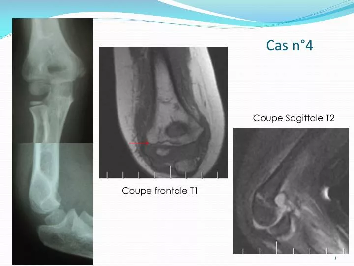
Cas n°4. Coupe Sagittale T2. Coupe frontale T1. Coupe Frontale T2 FS. Discussion. Milch H. Fractures of the external humeral condyle. JAMA 1956 ;160:641–6. Discussion. Stade I: déplacement inf à 2mm avec une surface articulaire intact

E N D
Cas n°4 Coupe Sagittale T2 Coupe frontale T1
Discussion Milch H. Fractures of the external humeral condyle. JAMA 1956;160:641–6.
Discussion Stade I: déplacement inf à 2mm avec une surface articulaire intact Stade II: déplacement entre 2-4mm discret déplacement articulaire Stade III: déplacement important avec rotation du fragment Stade I Stade II Stade III Jakob R, Fowles JV, Rang M, Kassab MT. Observations concerning fractures of the lateral humeral condyle in children. J Bone J Surg Br 1975;57:430–6.
Discussion Type I: trait non déplacé vu sur une seule incidence Type II: trait non déplacé vu sur deux incidences Type III: déplacement sup à 2mm sur deux incidences Type IV: séparation fragments Badelon O, Bensahel H, Mazda K, Vie P. Lateralhumeralcondylar fractures in children: a report of 47 cases. J PediatrOrthop 1988;8:31–4. Type I: trait ne traverse pas l’articulation Type II: trait atteint l’articulation Type III: déplacement du fragment Rutherford A. Fractures of the lateral humeral condyle in children. J Bone JointSurg Am 1985;67:851–6.
Echographie Ultrasonography for non-displaced and mini-displacedhumerallateral condyle fractures in children. Zhang JD, Chen H. Chin J Traumatol. 2008 Oct;11(5):297-300.
Tomodensitométrie Chapman VM, Grottkau BE, Albright M, Salamipour H, Jaramillo D.J Multidetectorcomputedtomography of pediatriclateralcondylar fractures. Comput AssistTomogr. 2005 Nov-Dec;29(6):842-6.
IRM • Kamegaya, Makoto M.D.; Shinohara, Yuhji M.D.et al. Assessmentof Stability in Children'sMinimallyDisplacedLateralHumeral Condyle fracture by MagneticResonance Imaging. J OrthopPediat1999;19:570-73 • T. PUDAS, T. HURME, K. MATTILA & E. SVEDSTRO¨MMagnetic Resonance Imaging in Pediatric Elbow Fractures; ACTA RADIOLOGICA. 2005. Departments of Radiology and Pediatric Surgery, University of Turku, Turku, Finland
Algorithme décisionnel Fracture peu déplacée du condyle latéral IRM Trait métaphysaire Trait métaphyso-épiphysaire Salter IV extra articulaire Fracture Salter IV Traitement orthopédique Fractures stables Traitement orthopédique +/- Embrochage percutané Traitement chirurgical