Download
1 / 52
550 likes | 864 Vues
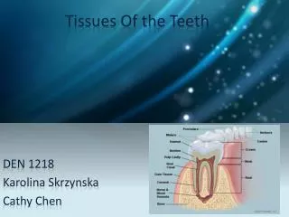
Tissue of the teeth. Dr Jamal Naim PhD in Orthodontics. Enamel. Introduction.

E N D
Tissue of the teeth Dr Jamal Naim PhD in Orthodontics Enamel
Introduction The current-day clinical practice of the dentistry involves the prevention of enamel demineralization, the promotion of enamel remineralization, the restoration of cavitated enamel where demineralization has become irreversible, and the diagnosis and treatment of developmental enamel malformations, which can be caused by environmental or genetic factors.
Introduction On a daily basis, dental health providers make diagnostic and treatment decisions that are influenced by their understanding of tooth formation. A systemic condition during tooth development, such as high fever, can produce a pattern of enamel defects in the dentition. Knowing the timing of tooth development permits estimates about the timing of the disturbance.
Introduction The process of enamel maturation continues following tooth eruption, so that erupted teeth can become less susceptible to decay over time. Mutations in the genes encoding enamel proteins lead to amelogenesis imperfecta, a collection of inherited diseases having enamel malformations as the predominant phenotype.
Enamel compositions Tooth enamel is unique among mineralized tissues because of its high mineral content. Enamel is made up of highly organized, tightly packed crystallites that comprise 87% percent of its volume and 95-96% of its weight.
Enamel compositions The inorganic Material (mineral structure): • is calcium hydroxyapatite [Ca10(PO4)6(OH)2] • contains impurities, such as carbonate substituting for phosphate in the crystals. Calcium hydroxyapatite can be synthesized from chemicals in the lab, but the shape, size, and organization of the crystals are always radically different from those of dental enamel.
Enamel compositions The organic materials are enamel proteins such as amelogenin (90%), ameloblastin and enamelin, (both 10%). The organic materials forms an organic network that transport minerals during the Enamel formation (amelogenesis) and determine the nature and direction of crystal growth.
Cross section (A) Neural crest cells
Ectoderm Mesoderm Basement membrane Basal cell layer
Basement membrane ECTODERM MESODERM ectomesenchymal cells
Bud stage Future dental papilla
cap stage Future dental papilla
Life History of ameloblasts It begins at the early Bell stage with: • Morphogenic Stage • Organizing stage • Formation stage (crystal lengthening) • Maturation stage (crystal thickening) • Protective stage • Desmolytic stage • Degenerative Stage
Life History of ameloblasts P Bud stage Cup stage Bell stage P P Bell stage Bell stage Root formation
Life History of ameloblasts • Morphogenic Stage: During embryonic development, cells covering the cranial neural crest (CNC) invade the underlying connective tissue and migrate into the maxillary and mandibular prominences. These migratory cells share characteristics of epithelial and connective tissues and are commonly referred to as “ectomesenchyme” .
Life History of ameloblasts Interactions between ectomesenchyme and the cells of the inner dental epithelium ultimately lead to the formation of two opposing sheets of columnar cells: ameloblasts and odontoblasts. Dentin forms on the side of the odontoblasts, and enamel forms on the side of the ameloblasts. The histology of tooth formation is classically divided into bud, cap, and bell stages.
Morphogenic stage Preameloblast Basement membrane Basement membrane
Life History of ameloblasts 2. Organizing stage: • Organizing of the preameloblasts in one cell layer • Changing of the cytomorphology • Migration of nucleus, centrions and Golgi regions from distal to proximal ends. This Stage begins directly after the initiation of dentin formation.
Morphogenic stage & Organizing stage Basement membrane
Life History of ameloblasts Secretory ameloblasts are tall, columnar cells with a proximally polarized nucleus and proximal and distal cell-cell junctions. Their length is about 40-50 micrometer and their thickness is about 5 micrometer.
Life History of ameloblasts 3. Formation (secretory) stage: prior to the onset of biomineralization, preameloblasts secrete enamel proteins on top of the predentin matrix. Some of the enamel proteins penetrate the predentin and are absorbed by odontoblasts.
Life History of ameloblasts Immediately following the initial secretion of enamel proteins, the basement membrane disappears, and ameloblast cell processes extend into irregularities on the predentin surface.
Formation stage reciprocal induction
Life History of ameloblasts Enamel crystallites are initiated within these irregularities, in close proximity to both the ameloblast cell membrane and collagen fibers protruding from the predentin. The ameloblastic processes appear to retreat back to the cell body, extending the incipient enamel crystallites as they go.
Life History of ameloblasts This fills in the irregular (villus) surface of dentin with enamel crystallites and converts it into the smooth, undulating surface of aprismatic enamel, which is perforated by odontoblastic processes.
Life History of ameloblasts Dentin and enamel are intimately linked at the dentino-enamel junction. The collagen-based organic matrix gives dentin its tensile strength and flexibility, and allows it to cushion the more brittle enamel covering.
Life History of ameloblasts After depositing the aprismatic enamel layer, secretory ameloblasts develop a specialized, cone-shaped Tomes’ process at their secretory (distal) ends. TP
Life History of ameloblasts Enamel Formation During the Secretory Stage: • Crystal Elongation Mineral deposition occurs primarily at a mineralization front very near to the ameloblast cell membrane. Enamel crystallites extend in length at extracellular growth sites a short distance away from the secretory faces of the ameloblast cell membrane.
Life History of ameloblasts During the secretory stage, enamel crystals do not grow continuously, but rather extend in increments (4 micron). Each increment represents the amount of crystal elongation that occurs in a single day, and is manifested structurally as prism cross-striations.
Life History of ameloblasts 4. Maturation stage: The ameloblasts become attached to the enamel matrix and exhibit microvilli at their distal ends. This alterations indicates the absorptive function of ameloblasts. In this stage the organic materials of enamel are being removed.
Life History of ameloblasts Ameloblast in the maturative stage
Life History of ameloblasts A significant feature of the maturation stage is that the pH of the fluid surrounding enamel crystals oscillates from less than 6 to 7.2. These pH fluctuations are similar to those the tooth will experience naturally, following its eruption into the oral cavity.
Life History of ameloblasts The developing enamel crystals are not structurally homogeneous. Crystals that are more susceptible to acid dissolution (i.e., those with high carbonate content) are selectively removed during the low pH part of the cycle. Therefore, during the maturation stage, an evolutionary process occurs in which relatively acid labile mineral is replaced by more acid- resistant apatite.
Life History of ameloblasts If the crown of a tooth become exposed to the oral cavity prematurely, such a tooth would be expected to decay rapidly due to the incomplete state of enamel maturation. This happens, when an unerupted third molar is transplanted into the socket of an extracted second molar. It should be treated with fluoride and sealant as soon as possible after the transplantation procedure.
Life History of ameloblasts At the end of the maturation stage, about 90 percent of the enamel volume is mineral, which contains less than 1 percent residual protein.
Life History of ameloblasts 5. Protective stage: After accomplishing of amelogenesis, the ameloblasts secrete or leave structure less material on enamel surface, known as primary enamel cuticle. The dental organ epithelium becomes reduced in thickness (reduced enamel epithelium) and functions as a protection against contact with connective tissue to inhibit cementum deposition or enamel resorption.
Life History of ameloblasts In this stage the composition of Enamel can be modified.
Life History of ameloblasts 6. Desmolytic stage: The reduced enamel epithelium seems to induce atrophy of the mesoderm which separate it from the oral epithelium. It leads to eruption.
Life History of ameloblasts 7. Degenerative Stage: As the tooth erupts in the mouth cavity, reduced enamel epithelium and oral epithelium fuse together. The new epithelium is called primary attachment epithelium. This degenerates and becomes replaced by oral epithelial cells forming the secondary attachment epithelium.