Download
1 / 47
560 likes | 1.02k Vues
Cardiac Arrhythmias in the SICU. Charles Hobson, MD MHA Surgical Critical Care NFSG VA Medical Center. Objectives. Review the etiology and recognition of common arrhythmias seen in the SICU. Review management of cardiac arrhythmias, with a focus on the relevant recent literature.

E N D
Cardiac Arrhythmias in the SICU Charles Hobson, MD MHA Surgical Critical Care NFSG VA Medical Center
Objectives • Review the etiology and recognition of common arrhythmias seen in the SICU. • Review management of cardiac arrhythmias, with a focus on the relevant recent literature.
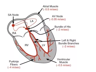
Normal Sinus Rhythm www.uptodate.com Implies normal sequence of conduction, originating in the sinus node and proceeding to the ventricles via the AV node and His-Purkinje system. EKG Characteristics: Regular narrow-complex rhythm Rate 60-100 bpm Each QRS complex is proceeded by a P wave P wave is upright in lead II & downgoing in lead aVR
Mechanisms of Arrhythmias Automaticity or Ectopic Foci Reentry / Conduction Block
Decreased Automaticity www.uptodate.com Sinus Bradycardia
Increased/Abnormal Automaticity Sinus tachycardia Ectopic atrial tachycardia www.uptodate.com Junctional tachycardia
Ectopic Foci and Beats QRS is slightly different but still narrow, indicating that conduction through the ventricle is relatively normal Atrial Escape Beats normal ("sinus") beats sinus node doesn't fire leading to a period of asystole (sick sinus syndrome) p-wave has different shape indicating it did not originate in the sinus node, but somewhere in the atria.
Ectopic Foci and Beats Paroxysmal Supraventricular Tachycardia (PSVT) • A single ectopic focus fires near the AV node, which then conducts normally to the ventricles (usually initiated by a PAC) • The rhythm is always REGULAR • Prolonged runs of PSVT may result in atrial fibrillation or atrial flutter • May be terminated by carotid massage • Treatment: carotid massage, adenosine, Ca++ channel blockers, ablation • Adenosine preferred in hypotension, previous IV B-blocker Note REGULAR rhythm in the tachycardia Rhythm usually begins with PAC
Ectopic Foci and Beats Multifocal Atrial Tachycardia (MAT) • Multiple ectopic foci fire in the atria, all of which are conducted normally to the ventricles • The rhythm is always IRREGULAR • P-waves of different morphologies (shapes) may be seen • Commonly seen in pulmonary disease, acute cardiorespiratory problems, and CHF • Treatment: • Ca++ channel blockers, beta blockers, but antiarrhythmic drugs are often ineffective • potassium, magnesium (McCord et al, Chest 1998), Note IRREGULAR rhythm in the tachycardia
Ectopic Foci and Beats Junctional Escape Beats QRS is slightly different but still narrow, indicating that conduction through the ventricle is relatively normal there is no p wave, indicating that it did not originate anywhere in the atria, but since the QRS complex is still thin and normal looking, we can conclude that the beat originated somewhere near the AV junction.
Ectopic Foci and Beats Ventricular Escape Beats “PVCs” QRS is wide and much different looking than the normal beats. This indicates that the beat originated somewhere in the ventricles. • no p wave, indicating that the beat did not originate anywhere in the atria • a "retrograde” p-wave may sometimes be seen on the right hand side of beats that originate in the ventricles, indicating that depolarization has spread back up through the atria from the ventricles
PVC's are Dangerous When: • They are frequent (> 30% of complexes) or are increasing in frequency • The come close to or on top of a preceding T-wave (R on T) • Three or more PVC's in a row (run of V-tach) • Any PVC in the setting of an acute MI • PVC's come from different foci ("multifocal" or "multiformed") • These may result in ventricular tachycardia or fibrillation. “R on T phenomenon” time Unconverted V-tach to V-fib sinus beats V-tach
Causes of Ectopic Foci and Beats • hypoxic myocardium - chronic pulmonary disease, pulmonary embolus • ischemic myocardium - acute MI, expanding MI, angina • sympathetic stimulation - nervousness, exercise, CHF, hyperthyroidism • drugs & electrolyte imbalances - antiarrhythmic drugs, hypokalemia, imbalances of calcium and magnesium • bradycardia- a slow HR predisposes one to arrhythmias • enlargement of the atria or ventricles producing stretch in pacemaker cells
The Reentry Mechanism of Ectopic Beats & Rhythms Electrical Impulse Cardiac Conduction Tissue Fast Conduction Path Slow Recovery Slow Conduction Path Fast Recovery • Tissues with these type of circuits may exist: • in the SA node, AV node, or any type of heart tissue • in a “macroscopic” structure such as an accessory pathway in WPW
The Reentry Mechanism of Ectopic Beats & Rhythms Premature Beat Impulse Cardiac Conduction Tissue Repolarizing Tissue (long refractory period) Fast Conduction Path Slow Recovery Slow Conduction Path Fast Recovery • 1. An arrhythmia is triggered by a premature beat • 2. The beat cannot gain entry into the fast conducting pathway because of its long refractory period and therefore travels down the slow conducting pathway only
The Reentry Mechanism of Ectopic Beats & Rhythms Cardiac Conduction Tissue Fast Conduction Path Slow Recovery Slow Conduction Path Fast Recovery • 3. The wave of excitation from the premature beat arrives at the distal end of the fast conducting pathway, which has now recovered and therefore travels retrograde (backwards) up the fast pathway
The Reentry Mechanism of Ectopic Beats & Rhythms Cardiac Conduction Tissue Fast Conduction Path Slow Recovery Slow Conduction Path Fast Recovery • 4. On arriving at the top of the fast pathway it finds the slow pathway has recovered and therefore the wave of excitation ‘re-enters’ the pathway and continues in a ‘circular’ movement. This creates the re-entry circuit
Reentrant Rhythms • AV nodal reentrant tachycardia (AVNRT) • Supraventricular tachycardia • AV reentrant tachycardia (AVRT) • Wolf – Parkinson – White syndrome • Atrial flutter • Ventricular tachycardia • Atrial fibrillation • Ventricular fibrillation
Reentry Circuits as Ectopic Foci and Arrhythmia Generators • Atrio-Ventricular Nodal Re-entry • supraventricular tachycardia • Ventricular Re-entry • ventricular tachycardia • ventricular fibrillation • Atrial Re-entry • atrial tachycardia • atrial fibrillation • atrial flutter • Atrio-Ventricular Re-entry • Wolf Parkinson White • supraventricular tachycardia
AV Nodal Reentrant Tachycardia Rate 100-270 Normal QRS Aberrancy possible • Acute Rx: • Vagal maneuvers • Adenosine 6-12 mg IV push – beware of pro-arrhythmia • Ca++ channel blockers
Atrial Flutter www.uptodate.com Atrial flutter is caused by a reentrant circuit in the wall of the atrium EKG Characteristics: Typical: “sawtooth” flutter waves at a rate of ~ 300 bpm Flutter waves have constant amplitude, duration, and morphology through the cardiac cycle There is usually either a 2:1 or 4:1 block at the AV node, resulting in ventricular rates of either 150 or 75 bpm
Dx and Rx of Flutter Unmasking of flutter waves with adenosine. • Acute Rx: • ventricular rate control can be difficult • AV nodal blockers prevent 1:1 conduction • Ibutilide 1-2mg rapid IV infusion – have paddles ready • Rapid pacing or low voltage DC cardioversion is effective • Anticoagulation as per atrial fibrillation
Ventricular Tachycardia Rate 100-20 Wide QRS Monomorphic vs Polymorphic • Beware: • Accelerated idioventricular rhythm. Rate below 150, stable hemodynamics, benign prognosis. • SVT with aberrancy. Look at the 12 lead – not just a rhythm strip • Monomorphic vs. Polymorphic (long QT, bradycardia, ischemia) • Rx: • Unstable – DC cardioversion • Stable monomorphic – Procainamide, Amiodarone • Stable polymorphic - treat underlying etiology
Atrial Fibrillation www.uptodate.com • Atrial fibrillation is caused by numerous waves of depolarization spreading throughout the atria, leading to an absence of coordinated atrial contraction. • Classified as: • Recurrent: when AF occurs on 2 or more occasions • Paroxysmal: episodes that generally last </= 7 days (most last <24h) • Persistent: AF that last >/=7 days • Permanent: paroxysmal or persistent AF with failure to cardiovert or not attempted
Dx and Rx of Atrial Fibrillation Absent P waves Irregularly irregular ventricular response • Acute Rx: • rate control not rhythm control – AFFIRM trial (NEJM 2002): • B-blockers, Ca++ channel blockers, digoxin, amiodarone • Ibutilide 1-2mg rapid IV infusion – have paddles ready • Oral propafenone or flecainide – beware pro-arrhythmia • Low voltage DC cardioversion • Anticoagulation as per atrial fibrillation • On the horizon: vernakalant, an atrial-selective Na and K channel blocker for conversion of short-duration atrial fibrillation
Ventricular Fibrillation www.uptodate.com Ventricular fibrillation is caused by numerous waves of depolarization spreading throughout the ventricles simultaneously, leading to disorganized ventricular contraction and immediate loss of cardiac function. EKG Characteristics: Absent P waves Disorganized electrical activity Deflections continuously change in shape, magnitude and direction
Rhythms Produced by Conduction Block • AV Block (relatively common) • 1st degree AV block • Type 1 2nd degree AV block • Type 2 2nd degree AV block • 3rd degree AV block • SA Block (relatively rare)
1st Degree AV Block The Alan E. Lindsay ECG Learning Center ; http://medstat.med.utah.edu/kw/ecg/ EKG Characteristics: Prolongation of the PR interval, which is constant All P waves are conducted Usually benign
2nd Degree AV Block Mobitz 1 (Wenckebach) EKG Characteristics: Progressive prolongation of the PR interval until a P wave is not conducted. As the PR interval prolongs, the RR interval actually shortens Usually benign unless associated with underlying pathology, i.e. MI
2nd Degree AV Block Mobitz 2 • EKG Characteristics: Constant PR interval with intermittent failure to conduct • Rhythm is dangerous as the block is lower in the conduction system • May cause syncope or may deteriorate into complete heart block • Causes: anterioseptal MI, fibrotic disease of the conduction system • Treatment: may require pacemaker in the case of fibrotic conduction system
3rd Degree (Complete) AV Block • EKG Characteristics: No relationship between P waves and QRS complexes • Constant PP intervals and RR intervals • May be caused by inferior MI and it’s presence worsens the prognosis • May cause syncopal symptoms, angina, or CFH • Treatment: usually requires pacemaker
Right Bundle Branch Block (RBBB) • Depolarization spreads from the left ventricle to the right ventricle. • This creates a second R-wave (R’) in V1, and a slurred S-wave in V5 - V6. • The T wave should be deflected opposite the terminal deflection of the QRS complex. This is known as appropriate T wave discordance with bundle branch block. A concordant T wave may suggest ischemia or myocardial infarction.
Left Bundle Branch Block (LBBB) • Depolarization enters the right side of the right ventricle first and simultaneously depolarizes the septum from right to left. • This creates a QS or rS complex in lead V1 and a monophasic or notched R wave in lead V6. • The T wave should be deflected opposite the terminal deflection of the QRS complex. This is known as appropriate T wave discordance with bundle branch block. A concordant T wave may suggest ischemia or myocardial infarction.
Class 1A agents: Procainamide, quinidine • Uses • Wide spectrum, but side effects limit usage • Quinidine : maintain sinus rhythms in atrial fibrillation and flutter and to prevent recurrent tachycardia and fibrillation • Procainamide: acute treatment of supraventricular and ventricular arrhythmias (no longer in production) • Side effects • Hypotension, reduced cardiac output • Proarrhythmia (generation of a new arrhythmia) eg. Torsades de Points (QT interval) • Dizziness, confusion, insomnia, seizure (high dose) • Gastrointestinal effects (common) • Lupus-like syndrome (esp. procainamide)
Class 1B agents: Lidocaine, phenytoin Uses acute : Ventricular tachycardia and fibrillation (esp. during ischemia) Not used in atrial arrhythmias or AV junctional arrhythmias Side effects Less proarrhythmic than Class 1A (less QT effect) CNS effects: dizziness, drowsiness
Class 1C agents: Flecainide, propafenone Uses Wide spectrum Used for supraventricular arrhythmias (fibrillation and flutter) Premature ventricular contractions (caused problems) Wolff-Parkenson-White syndrome Side effects Proarrhythmia and sudden death especially with chronic use (CAST study) Increase ventricular response to supraventricular arrhythmias CNS and gastrointestinal effects like other local anesthetics
Class II agents: Propranolol, esmolol Uses treating sinus and catecholamine dependent tachy arrhythmias converting reentrant arrhythmias in AV protecting the ventricles from high atrial rates (slow AV conduction) Side effects bronchospasm hypotension beware in partial AV block or ventricular failure
Class III agents: Amiodarone, sotalol, ibutilide Amiodarone Uses Very wide spectrum: effective for most arrhythmias Side effects: many serious that increase with time Pulmonary fibrosis Hepatic injury Increase LDL cholesterol Thyroid disease Photosensitivity May need to reduce the dose of digoxin and class 1 antiarrhythmics
Class III agents: Amiodarone, sotalol, ibutilide Sotalol Uses Wide spectrum: supraventricular and ventricular tachycardia Side effects Proarrhythmia, fatigue, insomnia
Class III agents: Amiodarone, sotalol, ibutilide Ibutilide Uses conversion of atrial fibrillation and flutter with rapid IV infusion Side effects Torsades de pointes
Class IV agents: Verapamil and diltiazem Uses control ventricular rate during supraventricular tachycardia convert supraventricular tachycardia (re-entry around AV) Side effects Caution when partial AV block is present. Can get asystole if β blocker is on board Caution when hypotension, decreased CO or sick sinus Some gastrointestinal problems
Additional agents Adenosine Administration rapid i.v. bolus, very short T1/2 (seconds) Cardiac effects Slows AV conduction Uses convert re-entrant supraventricular arrhythmias hypotension during surgery, diagnosis of CAD Magnesium treatment for tachycardia resulting from long QT
Additional agents Digoxin (cardiac glycosides) Mechanism enhances vagal activity, inhibits Na/K ATPase refractory period, slows AV conduction Uses treatment of atrial fibrillation and flutter Atropine Mechanism selective muscarinic antagonist Cardiac effects blocks vagal activity to speed AV conduction and increase HR Uses treat vagal bradycardia
Selected References: • ACC/AHA/ESC Practice Guidelines: • Supraventricular Arrhythmias – JACC 2003;42:1993-531. • Atrial Fibrillation – JACC 2006;48:854-906 • Ventricular Arrhythmias – JACC 2006;48:1064-1108 • Thanks, and questions?