Download
1 / 24
260 likes | 397 Vues
General Pathology. Cellular and Organ Pathology Disorders of Glycogen Degradation. Pathology of Calcification. Jaroslava Dušková Inst. Pathol. ,1st Med. Faculty, Charles Univ. Prague. Glycogen. linear and branched polymer cca 60 000 - D-glucose molecules monoparticles (beta) - muscle

E N D
General Pathology Cellular and Organ Pathology Disorders of Glycogen Degradation. Pathology of Calcification. Jaroslava Dušková Inst. Pathol. ,1st Med. Faculty, Charles Univ. Prague
Glycogen • linear and branched polymer • cca 60 000 - D-glucose molecules • monoparticles (beta) - muscle • complex particles (alpha) - hepatocyte
EnzymesInvolved in Glycogen Metabolism • g.-synthase - brancher • phophorylase kinase - debrancher • g-6-phosphatase • -glucosidase
Storage Diseases Def.: inborn errors of metabolism (mostly single gene abnormality) leading to an enzyme defect with subsequent accumulation of the substrate (& lack of the product) in tissues or organs„thesaurismoses“
Glycogenosis I – von Gierke E defect - gl-6 - phosphatase Organ damage - liver, kidney
Glycogenosis II – Pompe E -defect - alfa1,4-glycosidase Organ damage - heart
Clear Intracellular Vacuoles& adjunct techniques • accumulations of water neg. • lipides SUDAN, OILRED • polysaccharides PAS, A-PAS
Glycogen water soluble easily lost in long lasting water based fixative solutions
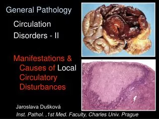
Calcification Def.: depositions of Ca (mostly phosphate salts) in tissues or organs Classification: dystrophic metastatic
Calcification - physiology • Matrix vesicles - osteoblasts- nidus calcification • Non collagen proteins - osteopontin, osteonektin, osteokalcin, Gla protein,sialoprotein • Alkalic phosphatase • Phospholipids • Collagen I • Hydroxyapatite
Pathology Conditions with Calcium Deposits Calcification • dystrophic • metastatic Calcinosis • localized • generalized Chondrocalcinosis-pseudogout
Calcification - Microscopy • Basophilic • Von Kossa - Ag impregnation • Alizarine red + • Tetracyclin fluorescence • Polarized light birefringence
Dystrophic Serum Ca level: normal Tissues/Organs status dystrophic changes (necrosis, scar…, low metab. turnover) Metastatic Serum Ca level: Tissues/Organs status normal, local alcalisation (acid secretion - urine, stomach juice, sweat…) Calcification
Dystrophic Serum Ca level: normal Tissues/Organs status dystrophic changes (necrosis, scar…, low metab. turnover) Metastatic Serum Ca level: Tissues/Organs status normal, local alcalisation (acid secretion - urine, stomach juice, sweat…) Calcification
Ca Salts in the Calcified Foci • Ca phophate Ca3(PO4)2 • Ca diphosphate (Ca2P2O7) • Hydroxyapatite (Ca5 (PO4)3.OH
Pathology Conditions with Calcium Deposits Calcification • dystrophic • metastatic Calcinosis • localized • generalized Chondrocalcinosis-pseudogout
Dystrophic Calcification • Frequent • necrotic tissue • connective tissue • vessels • kalkospherites • intracellular calcifiction of lysosoms Less frequent tendon, cartilage, elastics, bas. membranes
Calcinosis Forms - localised generalised Localisation– connective tissue, muscles
Chondrocalcinosis crystal deposit disease (pseudogout) pyrophophate and hydroxyapatite deposits localisation– synovial membrane, cortilage, bone
Calcium pyrophosphate deposition disease(Chondrocalcinosis, pseudogout) • Clinic: may simulate different diseases • Arthroscopy - chalky white deposits • Tophaceus deposits with crystalline aggregates • Crystals stain with von Kossa technique • Foreign body type multinucleated giant cell reaction • Deposits are in synovium, cartilage, bone