Download
1 / 13
230 likes | 988 Vues
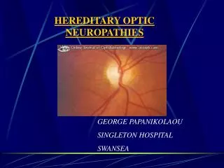
OPTIC NEUROPATHIES. 1. Clinical features. 2. Special investigations. 3. Optic neuritis. Retrobulbar neuritis Papillitis Neuroretinitis. 4. Anterior ischaemic optic neuropathy (AION). 5. Leber hereditary optic neuropathy. Signs of optic nerve dysfunction.

E N D
OPTIC NEUROPATHIES 1. Clinical features 2. Special investigations 3. Optic neuritis • Retrobulbar neuritis • Papillitis • Neuroretinitis 4. Anterior ischaemic optic neuropathy (AION) 5. Leber hereditary optic neuropathy
Signs of optic nerve dysfunction • Reduced visual acuity • Afferent pupillary • conduction defect • Dyschromatopsia • Diminished light • brightness sensitivity
Applied anatomy of afferent conduction defect Anatomical pathway Signs • Equal pupil size • Light reaction • - ipsilateral direct is absent or diminished • - consensual is normal • Near reflex is normal in both eyes • Total defect (no PL) = amaurotic pupil • Relative defect = Marcus Gunn pupil 3rd
Visual field defects Central scotoma Centrocaecal scotoma Altitudinal Nerve fibre bundle
Optic disc changes Normal Swelling • Papilloedema • Retrobulbar neuritis • Papillitis and neuroretinitis • Early compression • AION Optico-ciliary shunts Atrophy • Postneuritic • Optic nerve sheath meningioma • Compression • Occasionally optic nerve glioma • Hereditary optic atrophies
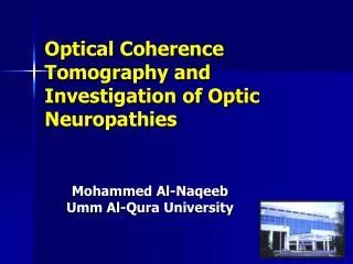
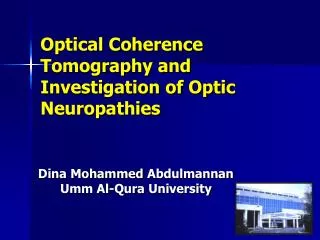
Special investigations MRI Visually evoked potential Assessment of electrical activity of visual cortex created by retinal stimulation Orbital fat-suppression techniques in T1-weighted images
Classification of optic neuritis Retrobulbar neuritis (normal disc) Papillitis (hyperaemia and oedema) Neuroretinitis (papillitis and macular star) • Demyelination - most common • Cat-scratch fever • Viral infections and immunization • in children (bilateral) • Sinus-related (ethmoiditis) • Lyme disease • Demyelination (uncommon) • Lyme disease • Syphilis • Syphilis
Non-arteritic AION Presentation • Age - 45-65 years • Altitudinal field defect • Eventually bilateral in 30% (give aspirin) Acute signs Late signs • Pale disc with diffuse or sectorial oedema • Resolution of oedema and haemorrhages • Few, small splinter-shaped haemorrhages • Optic atrophy and variable visual loss
FA in acute non-arteritic AION Increasing localized hyperfluorescence Localized hyperfluorescence Generalized hyperfluorescence
Superficial temporal arteritis Presentation • Age - 65-80 years • Scalp tenderness • Headache • Jaw claudication • Polymyalgia rheumatica • Superficial temporal arteritis • Acute visual loss Special investigations • ESR - often > 60, but normal in 20% • C-reactive protein - always raised • Temporal artery biopsy
Histology of giant cell arteritis • Granulomatous cell infiltration • High-magnification shows giant cells • Disruption of internal elastic lamina • Proliferation of intima • Occlusion of lumen
Arteritic AION • Affects about 25% of untreated patients with giant cell arteritis • Severe acute visual loss • Treatment - steroids to protect fellow eye • Bilateral in 65% if untreated • Pale disc with diffuse oedema • Few, small splinter-shaped haemorrhages • Subsequent optic atrophy
Leber hereditary optic neuropathy Maternal mitochondrial DNA mutations Presents • Typically in males - third decade • Occasionally in females - any age • Initially unilateral visual loss • Fellow eye involved within 2 months • Bilateral optic atrophy Signs • Disc hyperaemia and dilated capillaries • (telangiectatic microangiopathy) • Vascular tortuosity • Swelling of peripapillary nerve fibre layer • Subsequent bilateral optic atrophy