Download
1 / 48
500 likes | 727 Vues
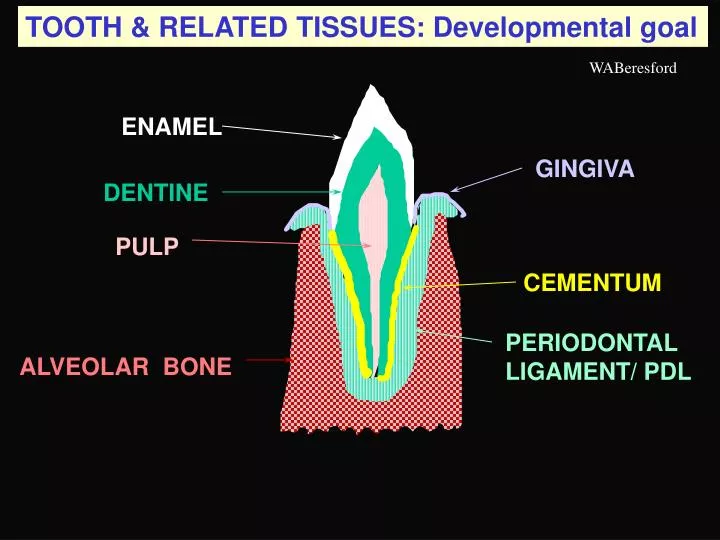
TOOTH & RELATED TISSUES: Developmental goal. WABeresford. ENAMEL. GINGIVA. DENTINE. PULP. CEMENTUM. PERIODONTAL LIGAMENT/ PDL. ALVEOLAR BONE. What has to be controlled. Whether teeth are to form ( turtles & birds have beaks). Number of teeth. Position of teeth. Shapes of teeth.

E N D
TOOTH & RELATED TISSUES: Developmental goal WABeresford ENAMEL GINGIVA DENTINE PULP CEMENTUM PERIODONTAL LIGAMENT/ PDL ALVEOLAR BONE
What has to be controlled Whether teeth are to form (turtles & birds have beaks) Number of teeth Position of teeth Shapes of teeth Crown formed before root Four tooth tissues in sequence Organize surroundings Fasten tooth to surroundings Times of eruption Shedding of teeth Successional teeth Protect enamel
MECHANISMS OF DEVELOPMENT 10 Ectodermal laminae can be used: At discrete points only, for making dental organs of tooth germs. The remainder of the lamina then breaks up and disappears, but a few ectodermal cells may remain as epithelial rests or pearls LAMINA DENTAL ORGAN
DENTAL LAMINA Dental lamina the line of thickened ectoderm Mesenchyme Oral ectoderm
TOOTH DEVELOPMENT BRAIN TOOTH BUD TONGUE DENTAL LAMINA from which DENTAL ORGANS (tooth germs) form TOOTH
FIRST DENTAL ARCHES FROM BELOW UPPER LIP TECTAL RIDGE (future Gum) Left Maxillary arch - starts as a dental lamina in oral ectoderm overlying tectal-ridge mesenchyme LATERAL PALATINE SHELF
DENTAL LAMINA Dental lamina line of thickened ectoderm Mesenchyme Oral ectoderm
MECHANISMS OF DEVELOPMENT 10 Ectodermal laminae can be used: At discrete points only, for making dental organs of tooth germs. The remainder of the lamina then breaks up and disappears, but a few ectodermal cells may remain as epithelial rests or pearls REST LAMINA DENTAL ORGAN
DENTAL LAMINA: Two meanings LAMINA Dental lamina first refers to the thickening in the ectoderm along the tectal ridge. From this a secondary dental lamina grows down into the mesenchyme. At intervals along this deep lamina, dental organs (tooth buds) form. LAMINA As the dental organs are established, the original surface lamina reverts to oral lining ectoderm (differentiating into gingival epithelium) & the secondary lamina starts to disintegrate, leaving the first dental organ & a successional lamina for the second tooth bud
DENTAL LAMINA Dental lamina first sense - the line of thickened ectoderm Mesenchyme Dental laminasecond sense - the individual downgrowths from the ectoderm Oral ectoderm Next step - formation of a BUD from the dental lamina
DENTAL LAMINA Oral ectoderm Mesenchyme Dental lamina line of thickened ectoderm What is to be controlled: Whether teeth are to form Lamina - yes Line of teeth Line of lamina in arch Position of teeth Siting of tooth buds Number of teeth # of dental organs Successors of teeth to be shed Successional laminae & buds
JAW & TOOTH DEVELOPMENT early arch BONE starting BUCCAL PLATE DENTAL LAMINA BONE starting LINGUAL PLATE 10 TOOTH GERM 20 Successional TOOTH GERM WALLS OF BONY TROUGH OF DEVELOPING MANDIBLE SYMPHYSEAL CARTILAGE
JAW & TOOTH DEVELOPMENT processes DENTAL LAMINA will grow back to form germs for 3 permanent molars (5th e m) BONE 10 TOOTH GERM 20 Successional TOOTH GERM on lingual side of 10 PLANTING POTATOES Bone creates the trench; tooth buds are the spuds
TOOTH PRIMORDIUM/GERM DENTAL ORGAN DENTAL LAMINA MESENCHYME DENTAL PAPILLA DENTAL SAC/FOLLICLE ALVEOLAR BONE
TOOTH TISSUES: Sources ENAMEL DENTAL ORGAN DENTAL LAMINA DENTINE PULP MESENCHYME CEMENTUM PDL DENTAL PAPILLA DENTAL SAC/FOLLICLE A BONE ALVEOLAR BONE
TOOTH TISSUES: Cell Sources TOOTH DENTAL LAMINA DENTAL ORGAN ENAMEL Ameloblasts DENTINE DENTAL PAPILLA Odontoblasts PULP CT cells CEMENTUM DENTAL SAC/FOLLICLE Cementoblasts PDL Fibroblasts ALVEOLAR BONE A BONE Osteoblasts & ‘clasts Crest
TOOTH TISSUES: Sources TOOTH DENTAL LAMINA DENTAL ORGAN ENAMEL DENTINE DENTAL PAPILLA PULP CEMENTUM DENTAL SAC/FOLLICLE PDL LAMINA DURA ALVEOLAR BONE SUPPORTING BONE Plate Spongy bone
TOOTH GERM Outer dental epithelium DENTAL LAMINA Stellate reticulum Stratum intermedium DENTAL PAPILLA Inner dental epithelium DENTAL SAC/FOLLICLE
TOOTH GERM: context Dental lamina Oral ectoderm Vestibular lamina Mesenchyme
Oral ectoderm Dental lamina Vestibular lamina will grow down and split on the tooth’s labial/buccal side creating alveolar & labial/buccal mucosae
DENTAL LAMINA Dental lamina first sense - the line of thickened ectoderm Mesenchyme Dental lamina second sense - the individual downgrowths from the ectoderm Oral ectoderm Next step - formation of a BUD from the dental lamina
DENTAL LAMINA: Bud stage Formation of a BUD from the dental lamina Odontogenic mesenchyme Next step - formation of a CAP from the bud
DENTAL ORGAN: Cap, becoming Bell stage Outer dental epithelium DENTAL LAMINA Enamel knot Stellate reticulum Stratum intermedium Basal lamina Inner dental epithelium Cap determines: position, type, & size of tooth
TOOTH GERM: next steps Outer dental epithelium approaches inner DENTAL LAMINA upper part degenerates lower forms 2nd bud Stellate reticulum reduces over cusp Knot cells signal to papilla Stratum intermedium DENTAL PAPILLA Inner & outer dental epithelia join to form cervical loop DENTAL SAC/FOLLICLE
TOOTH GERM: result & next steps Outer dental epithelium collapsing down DENTAL LAMINA upper degenerates lower forms 2nd bud Stellate reticulum reducing over cusp Knot cells signal to papilla outermost papilla cells have become Odontoblasts DENTAL PAPILLA becoming pulp Recruitment site Cervical loop defines extent of crown to crown base; then it starts the root sheath DENTAL SAC quiescent Ingrowing pulp vessels
TOOTH GERM: result & next steps Dentine is formed first as predentine. It will signal to inner dental epithelialcells to become ameloblasts DENTAL LAMINA upper degenerates lower forms 2nd bud Inner dental epithelium Stellate reticulum moving “apically” cuspDentine formed by Odontoblasts DENTAL PAPILLA becoming pulp Recruitment site Cervical loop moving apically todefine extent of crown DENTAL SAC still quiescent Ingrowing pulp vessels
Capillaries now close to synthesizing ameloblasts Ameloblasts Stellate reticulum cusp enamelformed byameloblasts Dentine DENTAL PAPILLA becoming pulp Cervical loop moving apically todefine extent of crown DENTAL SAC still quiescent 2nd TOOTH GERM: all crown-forming elements present
ROOT FORMATION REDUCED DENTAL EPITHELIUM ENAMEL CROWN DENTINE PULP Root sheath breaks up, allowing sac mesenchymalcells to contact root dentine HERTWIG’S ROOT SHEATH Odontoblast recruitment site Epithelial diaphragm
FURTHER ROOT FORMATION REDUCED DENTAL EPITHELIUMwill fuse with gingiva Root sheath breaks up, allowing sac mesenchymalcells to contact root dentine ENAMEL matures DENTINE deepens PULP differentiates Other sac mesenchymalcells construct PDL & some alveolar bone HERTWIG’S ROOT SHEATH grows to lengthen root Fibroblasts Odontoblast recruitment site Epithelial diaphragm
TWO STORIES: crown & root Crown details continue Root details in Eruption.ppt
CHANGES IN DENTAL/ENAMEL ORGAN I Capillaries Ameloblasts Stellate reticulum Starting at the cusp the dental organ reduces to two layers: active ameloblasts & a narrow layer of compacted outer epithelium, stellate-reticulum cells & stratum intermedium Stratum intermedium Cervical loopmoving apically todefine extent of crown & pinches in
CHANGES IN DENTAL/ ENAMEL ORGAN II cusp enamelformed byameloblasts First reduction in enamel epithelium:active ameloblasts& compacted outer epithelium, stellate-reticulum cells & stratum intermedium Dentine PULP from Dental Papilla remaining Stellate reticulum DENTAL PAPILLA still becoming pulp & odontoblasts Cervical loop: inner & outer epithelium DENTAL SAC still quiescent
CHANGES IN DENTAL/ ENAMEL ORGAN III Ameloblasts willfinish full thickness of cusp enamel & reduce in height Second reduction in enamel epithelium:retired ameloblasts& compacted outer epithelium, stellate-reticulum cells & stratum intermedium Dentine widens DENTAL PAPILLA becomes pulp process proceeds downs Stellate reticulum follows Cervical loop down then stops: Crown defined Odontoblast recruitment site Cervical loop:
ROOT FORMATION ENAMEL REDUCED DENTAL EPITHELIUM CROWN DENTINE Where Stellate reticulum stopped, the cervical loop continued to grow down, but as PULP HERTWIG’S ROOT SHEATH & its ROOT Odontoblast recruitment site Epithelial diaphragm
ROOT FORMATION: Coronal consequences GINGIVAL EPITHELIUM Connective tissue broken down REDUCED DENTAL EPITHELIUMwill fuse with gingiva REDUCED DENTAL EPITHELIUMprotects enamel As root lengthens crown is pushed up - Pre-EMERGENCE HERTWIG’S ROOT SHEATH grows to lengthen root
FURTHER CORONAL EVENTS GINGIVAL EPITHELIUM fusing with REDUCED DENTAL EPITHELIUM still protecting enamel ENAMEL Root lengthening not shown
TOOTH EMERGENCE CUTICLE will wear away GINGIVAL EPITHELIUM still fusing with REDUCED DENTAL EPITHELIUM ENAMEL
EVENTS WHERE EPITHELIA MEET? CUTICLE will wear away GINGIVAL EPITHELIUM still fusing with Which reduced-epithelial cells cells join the gingiva, which contribute to the cuticle, & which die, is unclear; along with the mechanisms determining fate REDUCED DENTAL EPITHELIUM
ORGANIC ENAMEL SURFACE CUTICLE will wear away PELLICLE of glycoproteins etc is acquired later from oral sources
DENTAL LAMINA Dental lamina first sense - the line of thickened ectoderm Mesenchyme Dental lamina second sense - the individual downgrowths from the ectoderm Oral ectoderm Next step - formation of a BUD from the dental lamina
DENTAL LAMINA: Bud stage Formation of a BUD from the dental lamina Odontogenic mesenchyme Next step - formation of a CAP from the bud
REPEATED BACK-&-FORTH SIGNALING I Oral ectoderm to mesenchyme Dental lamina Odontogenic mesenchyme Early signaling center SIGNALS: FGFs, BMPs, Wnt, etc Condensed dental mesenchyme
REITERATIVE SIGNALING II Enamel knot Inner enamel epithelium Knot cellssignal to epithelial cells to proliferate, extending inner epithelium, & creating Bell-shaped dental/enamel organ
REITERATIVE SIGNALING III Knot cells signal to papilla outermost papilla cells become Odontoblasts
REITERATIVE SIGNALING IV outermost papilla cells have become Odontoblasts Odontoblasts make dentine, which signals inner epithelial cells to beome ameloblasts
REITERATIVE SIGNALING V REDUCED DENTAL EPITHELIUM ENAMEL DENTINE Root sheath breaks up & lifts, allowing sac mesenchymalcells to contact root dentine Dentine &/or Epithelial root sheathinduces mesenchymal cellsto become cementoblasts PULP Odontoblast recruitment site byroot sheath: pulp signaling
What has to be controlled:structures & mechanisms W hether teeth are to form (turtles & birds have beaks) Dental lamina Number of teeth # of buds siting of buds Position of teeth shape of dental organ Shape(s) of teeth Crown formed before root start with cusp tissues root sheath for root control Four tooth tissues in sequence mesenchyme for three, ectoderm for one tissue Organize surroundings alveolar bony trough & dental sac mesenchyme Fasten tooth to surroundings dental sac mesenchyme Times of eruption ? & sequence of formation Shedding of teeth Use bone remodeling cells on root Successional teeth Successional lamina & bud
To be controlled:structures & mechanisms W hether teeth are to form (turtles & birds beaks) Dental lamina Number of teeth # of buds Position of teeth siting of buds Shape(s) of teeth shape of dental organ start with cusp tissues root sheath for root control Crown formed before root mesenchyme for three, ectoderm for one tissue 4 tooth tissues in sequence alveolar bony trough & dental sac mesenchyme Organize surroundings Fasten tooth to surroundings dental sac mesenchyme Times of eruption ? & sequence of formation Shedding of teeth Use bone remodeling cells on root Successional teeth Successional lamina & bud Protect enamel Reduced enamel epithelium