Download
1 / 36
360 likes | 581 Vues
Chapter 13: Recognizing Different Sports Injuries. No matter how much time is spent on injury prevention, sooner or later an injury occurs Either acute or chronic in nature Acute injuries Result of trauma Chronic Caused by repetitive, overuse activities. Acute Traumatic Injuries. Fractures.

E N D
No matter how much time is spent on injury prevention, sooner or later an injury occurs • Either acute or chronic in nature • Acute injuries • Result of trauma • Chronic • Caused by repetitive, overuse activities
Fractures • Result of extreme stress and strain on bone • Anatomical Characteristics • Dense connective tissue matrix • Outer compact tissue • Inner porous cancellous bone including Haversian canals
Gross Structures • Diaphysis -shaft - hollow and cylindrical - covered by compact bone • Epiphysis - composed of cancellous bone and has hyaline cartilage covering • Periosteum - dense, white fibrous covering which penetrates bone via Sharpey’ fibers • - contains blood vessels and osteoblasts
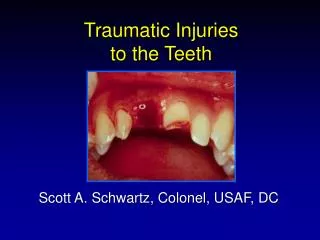
Acute bone fractures – • partial or complete disruption that can be either closed or open (through skin) • serious musculoskeletal condition • presents with deformity, point tenderness, swelling and pain on active and passive motion
Load Characteristics • Bones can be stressed or loaded to fail by tension, compression, bending, twisting and shearing • Either occur singularly or in combination • Amount of load also impacts the nature of the fracture • More force results in a more complex fracture • While force goes into fracturing the bone, energy and force is also absorbed by adjacent soft tissues • Some bones will require more force than others
Healing of a Fracture • Generally require immobilization for some period • Approx. 6 weeks for bones of arms and legs • 3 weeks for bones of hands and feet • Fracture healing requires osteoblast activity to lay down bone and form callus • Following cast removal, normal stresses and strains will aid in healing and remodeling process • Osteoclasts will be called on to assist in re-shaping of bone in response to normal stress
Stress fractures • No specific cause but with a number of possible causes • Overload due to muscle contraction, altered stress distribution due to muscle fatigue, changes in surface, rhythmic repetitive stress vibrations • Begins with a dull ache and progressively becomes worse over time • Initially pain during activity and then progresses to pain following activity • Early detection is difficult, bone scan is useful, x-ray is effective after several weeks • Due to osteoblastic activity • If suspected – stop activity for 14 days • Generally does not require casting
Dislocations and Subluxations • Dislocation • At least one bone in a joint is forced completely out of normal and proper alignment • High level of incidence in fingers, elbow and shoulder • Subluxation • Partial dislocations causing incomplete separation of two bones • Often occur in shoulder and females (patella) • S&S of dislocations • Deformity – almost always present • Occasionally obscured by heavy musculature = requires palpation to determine normal contours
Other factors associated with dislocations - 1) loss of limb function, 2) swelling and point tenderness • Additional concerns • Avulsion fractures • Growth plate separation • “Once a dislocation, always a dislocation” • Treatment • Dislocations (particularly first time) should always be considered and treated as a fracture until ruled out • X-ray is the only absolute diagnostic technique • Return to play often determined by extent of soft tissue damage
Ligament Sprains • Sprain • Damage to a ligament (provides support to a joint) • Synovial joint characteristics • 2 or more bones • Capsule or ligaments • Capsule is lined with synovial membrane • Hyaline cartilage • Joint cavity with synovial fluid • Blood and nerve supply with muscles crossing joint • Mechanoreceptors within joint structures provide feedback relative to position
Some joints will have meniscus (thick fibrocartilage) for shock absorption and stability • Ligaments • Thickened portions of the capsule or totally separate bands • Dictates partially the motions of the joint
Sprains • Result of traumatic joint twist that causes stretching or tearing of connective tissue • Graded based on the severity of injury • Grading System • Grade I - some pain, minimal loss of function, no abnormal motion, and mild point tenderness, slight swelling and joint stiffness • Grade II - pain, moderate loss of function, swelling, and instability, some tearing of ligament fibers and joint instability • Grade III - extremely painful, inevitable loss of function, severe instability and swelling, and may also represent subluxation
Restoration of joint stability is difficult with grade I and II injuries • Must rely on other structures around the joint • Rely heavily on muscles surrounding joint • Ligament has been stretched/partially torn causing development of inelastic scar • Ligament will not regain original tension • Increased muscle tension due to strength training will improve joint stability
Contusions • Result of sudden blow to body • Can be both deep and superficial • Hematoma results from blood and lymph flow into surrounding tissue • Minor bleeding results in discoloration of skin • May be painful to the touch and with active movement • Must be cautious and aware of more severe injuries associated with repeated blows • Calcium deposits may form with fibers of soft tissue • Myositis ossificans
Prevention relies on protection and padding • Particularly when dealing with myositis ossificans • Protection and rest may allow for calcium re-absorption • Surgery would not be necessary to remove • Quadriceps and biceps are very susceptible to developing myositis ossificans
Muscle Strains and Injuries • Causes • Stretch, tear or rip to muscle or adjacent tissue • Muscle Strain Grades • Grade I - some fibers have been stretched or actually torn resulting in tenderness and pain on active ROM, movement painful but full range present • Grade II - number of fibers have been torn and active contraction is painful, usually a depression or divot is palpable, some swelling and discoloration result
Grade III- Complete rupture of muscle or musculotendinous junction, significant impairment, with initially a great deal of pain that diminishes due to nerve damage • Tendon ruptures • Large tendon ruptures will require surgery • Rehabilitation • Lengthy process regardless of severity • Will generally require 6-8 weeks • Return to activity too soon may result in re-injury
Muscle Cramps and Spasms • Painful involuntary contraction • Attributed to dehydration/electrolyte imbalance • May lead to muscle or tendon injuries • Muscle Guarding • Following injury, muscles within an effected area contract to splint the area in an effort to minimize pain through limitation of motion • Involuntary muscle contraction in response to pain following injury • Not spasm which would indicate increased tone due to upper motor neuron lesion in the brain
Muscle Soreness • Overexertion in strenuous exercise resulting in muscular pain • Generally occurs following participation in activity that individual is unaccustomed with • Two types of soreness • Acute-onset muscle soreness - accompanies fatigue, and is transient muscle pain experienced immediately after exercise • Delayed-onset muscle soreness (DOMS) - pain that occurs 24-48 hours following activity that gradually subsides (pain free 3-4 days later) • Potentially caused by slight microtrauma to muscle or connective tissue structures
Prevents muscle soreness through gradual build-up of intensity • Treat with static or PNF stretching and ice application within 48-72 hours of insult
Nerve Injuries • Two main causes of injury • Compression and tension • May be acute or chronic • Causes pain and can result in a host of sensory responses (pinch, burn, tingle, muscle weakness, radiating pain) • Injuries can range from minor to severe and life-altering • Healing process is very slow and long-term • Optimal environment is critical • CNS vs. PNS repair
Chronic Overuse Injuries • Importance of Inflammation in Healing • Essential part of healing process • Must occur following tissue damage to initiate healing • Signs and Symptoms • Pain, redness, swelling, loss of function, and warmth • If source of irritation is not removed then inflammatory process becomes chronic
Tendinitis • Most common overuse problem in sports • Gradual onset, with diffuse tenderness due to repeated microtrauma and degenerative changes • Obvious signs of swelling and pain • May also experience crepitus (due to chemical products of inflammation) • Key for treatment is rest and removal of causal factors • Work to maintain fitness but avoid activities that aggravate condition
Tenosynovitis • Inflammation of synovial sheath • In acute case - rapid onset, crepitus, and diffuse swelling • Chronic cases result in thickening of tendon with pain and crepitus • Often develops in long flexor tendons of fingers • Treatment is similar to that of tendinitis • NSAID’s may also be of some assistance
Bursitis • Bursa • Fluid filled sac that develops in area of friction • Sudden irritation can cause acute bursitis, while overuse and constant external compression can cause chronic bursitis • Results in increased fluid production, causing increases in pressure due to limited space around anatomical structures • Signs and symptoms include swelling, pain, and some loss of function • Three most commonly irritated bursae • Subacromial, olecranon, and prepatellar bursa
Osteoarthritis • Wearing away of hyaline cartilage as a result of normal use • Changes in joint mechanics lead joint degeneration (the result of repeated trauma to tissue involved) • May be the result of direct blow, pressure of carrying and lifting heavy loads, or repeated trauma from an activity such as running or cycling • Commonly affects weight-bearing joints but can also impact shoulders and cervical spine
Symptoms include pain (as the result of friction), stiffness, prominent upon rising in the morning, localized tenderness, creaking, grating, and often localized to one side of the joint or generalized joint pain
Myofascial Trigger Points • Develop due to mechanical stress • Either acute strain or static postural positions producing constant tension in muscle • Typically occur in neck, and upper and lower back • Signs and Symptoms • Pain with palpation, with predictable pattern of referred pain which may also limit motion • Pain may increase with active and passive motion of involved muscle
Importance of the Healing Process Following Injury • Coach must possess understanding of both sequence and time frame for various phases of the healing process • Interference with healing process will delay return to full activity • Work to create optimal healing environment • Little can be done to speed the process, while much can be done to impede it
Inflammatory Response Phase • Begins immediately following injury – critically important • Without the inflammatory phase the other phases will not occur • Phagocytosis occurs to clean the injured area • Chemical mediators are released to facilitate healing • Symptomatically presents with the following • Redness, swelling, warmth, tenderness and loss of function • Stage lasts 2-4 days following injury
Fibroblastic Repair Phase • Proliferative and regenerative activity occurs resulting in scar formation (fibroplasia) • Occurs within initial hours of injury and continues up to 4-6 weeks • S&S of inflammatory phase subside • Athlete will still experience some tenderness and pain with motion • With increasing development of the scar, complaints of pain and tenderness will decrease
Maturation-Remodeling Phase • Long-term process • Re-alignment of scar tissue according to tensile forces acting on tissue • Re-align to position of maximum efficiency (parallel to lines of tension) • Tissue gradually resumes normal appearance and function • After 3 weeks • Firm, strong, contracted, nonvascular scar exists • Maturation may take several years to be totally complete