Download
1 / 70
700 likes | 808 Vues
Chapter 1 Musculoskeletal Tissue. Tissues of the body. Epithelial Nervous Connective Muscle. Epithelial tissue. Two forms Membranous Forms such structures as the outer layer of the skin, the inner lining of the body cavities and lumina, and the covering of visceral organs Glandular

E N D
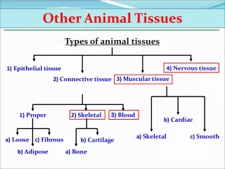
Tissues of the body • Epithelial • Nervous • Connective • Muscle
Epithelial tissue • Two forms • Membranous • Forms such structures as the outer layer of the skin, the inner lining of the body cavities and lumina, and the covering of visceral organs • Glandular • Specialized tissue that forms the secretory portion of glands
Nervous tissue • Helps coordinate movements via a complex motor control system of pre-structured motor programs and a distributed network of reflex pathways mediated throughout the CNS
Connective tissue • Found throughout the body • Divided into subtypes according to the matrix that binds the cells • Includes bone, cartilage, tendons, ligaments, and blood tissue
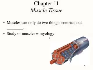
Muscle tissue • Responsible for the movement of materials through the body, the movement of one part of the body with respect to another, and locomotion • Three types: • Smooth • Cardiac • Skeletal
Connective Tissue • The primary types of connective tissue cells are: • Macrophages, which function as phagocytes to clean up debris • Mast cells, which release chemicals associated with inflammation • Fibroblasts, which are the principal cells of connective tissue
Connective Tissue Proper • 1.Loose connective tissue • 2.Dense regular connective tissue • 3.Dense irregular connective tissue • 4.Elastic connective tissue • 5.Reticular connective tissue • 6. Adipose connective tissue
Cartilage and bone tissue • 1.Hyaline cartilage • 2.Fibrocartilage • 3. Elastic cartilage
Collagen and Elastin • Collagen and elastin are vital constituents of the musculoskeletal system • Collagen • Maintains the structural integrity of various tissues • Provides tensile strength to tissues • Elastin • Provides the tissues in which it is situated with elastic properties
Collagen and Elastin • Collagenous and elastic fibers are sparse and irregularly arranged in loose connective tissue, but are tightly packed in dense connective tissue • Fascia is an example of loose connective tissue • Tendons and ligaments are examples of dense regular connective tissue
Tendons • Cordlike structures that function to attach muscle to bone and to transmit the forces generated by muscles to bone in order to achieve movement or stability of the body in space
Tendons • The fascicles of tendons are held together by loose connective tissue called endotenon • Endotenon contains blood vessels, lymphatics and nerves, and permits longitudinal movements of individual fascicles when tensile forces are applied to the structure • The connective tissue surrounding groups of fascicles and/or the entire structure is called the epitenon
Myotendinous junction (MTJ). • The site where the muscle and tendon meet • Very vulnerable to tensile failure, especially biceps and triceps brachii, the rotator cuff muscles, the flexor pollicis longus, the peroneus longus, the medial head of the gastrocnemius, the rectus femoris, the adductor longus, the iliopsoas, the pectoralis major, the semimembranosus, and the whole hamstrings group
Skeletal ligaments • Skeletal ligaments are fibrous bands of dense connective tissue that connect bones across joints • Contribute to the stability of joint function by preventing excessive motion • Act as guides to direct motion • Provide proprioceptive information for joint function
Ligament pathology • Ligament injuries are graded according to severity • First-degree (mild) • Second-degree (moderate) • Third degree (complete)
Ligament pathology • First-degree sprain • Minimal loss of structural integrity • Little or no swelling • Minimal bruising • Minimal functional loss • Early return to training
Ligament pathology • Second-degree sprain • Significant structural weakening • Some abnormal motion • More bruising and swelling • Tendency for recurrence • Need protection from risk of further injury • May need modified immobilization
Ligament pathology • Third-degree sprain • Loss of structural integrity • Marked abnormal motion • Significant bruising • Needs prolonged protection • Surgery may be considered • Permanent functional instability a possibility
Bone • A highly-vascular form of connective tissue • Composed of collagen, calcium phosphate, water, amorphous proteins, and cells • The collagen of bone is produced in the same manner as that of ligament and tendon, but by a different cell, the osteoblast
Bone • There are 206 bones in the human skeleton • 177 of these bones are involved in voluntary movement • 29 of these bones are immobile
Bone • The function of bone is to: • Provide support • Enhance leverage • Protect vital structures • Provide attachments for both tendons and ligaments • Store minerals, particularly calcium.
Axial Skeleton Skull Spinal Column Sternum Ribs Appendicular Skeleton Upper Extremity Lower Extremity Two Components of the Skeleton
Major Bone Types • Long • Short • Flat • Irregular
Long Bones Characteristics • Possess a cylindrical shaft and medullary canal • Relatively broad ends • Thick walled shaft
Upper Extremity Clavicle Humerus Ulna Radius Metacarpals Phalanges Lower Extremity Femur Tibia Fibula Metatarsals Phalanges Long Bones of Skeleton
Short Bones • Relatively short, compact and solid structures
Upper Extremity Carpals (wrist) Lower Extremity Tarsals (ankle) Short Bones of Skeleton
Flat Bones Examples: • Sternum • Scapulae • Ribs • Pelvic bones • Patellas
Irregular Bones Examples: • Bones of Spinal Column • The 24 vertebrae • Sacrum • Coccyx
Characteristics of Bone Epiphyses • Each bone has two epiphysis, which are layers of cartilage at the ends of the bones • The presence of an epiphysis indicates incomplete bone growth Articulation • Connection point of bones (joint) • Type of articulation helps determine the type and amount of motion possible
Pathology of bone • Osteoporosis • Maybe primary or secondary • Osteomalacia • Characterized by incomplete mineralization of normal osteoid tissue • Osteomyelitis • An acute or chronic inflammatory process of the bone and its marrow secondary to infection • Paget’s disease (osteitis deformans) • An osteometabolic disorder
Cartilage Tissue • Cartilage tissue consists of cartilage cells called chondrocytes • Chondrocytes are specialized cells that are responsible for the development of cartilage, and the maintenance of the extracellular matrix • Cartilage tissue exists in three forms: • Hyaline • Elastic • Fibrocartilage
Hyaline cartilage • Covers the ends of long bones and, along with the synovial fluid that bathes it, provides a smooth and almost frictionless articulating surface • The most abundant cartilage within the body
Elastic cartilage • A very specialized connective tissue, primarily found in locations such as the outer ear, and portions of the larynx
Fibrocartilage • Fibrocartilage functions as a shock absorber in both weight bearing, and non-weight bearing joints • Examples include the symphysis pubis, the intervertebral disc, and the menisci of the knee
Pathology of cartilage • Osteoarthritis • Can be primary or secondary • Osteochondritis dissecans • And osteochondral fracture
Joints • Joints are regions where bones are capped and surrounded by connective tissues that hold the bones together and determine the type and degree of movement between them • Joints may be classified as diarthrosis, which permit free bone movement and synarthrosis, in which very limited or no motion occurs
Joint Classifications • Synarthrodial (immovable) • Amphiarthrodial (slightly movable) • Syndesmosis • Synchondrosis • Diarthrodial (freely movable)
Diarthrosis • This type of joint generally unites long bones and has great mobility. Examples include but are not limited to the hip, knee and shoulder and elbow joints
Diarthrodial Joint Characteristics • Articular cavity present • Joint encased within ligamentous capsule • Capsule lined with synovial membrane • Secretion of synovial fluid, which lubricates the joint • Smooth articular surfaces, which are covered with cartilage
Diarthrodial Joint Classifications • Ball and socket (spheroidal; enthrodial) • Hinge (ginglymoid) • Pivot (trochoid) • Condyloid (ellipsoidal) • Irregular (arthrodial: plane) • Sellar (saddle)
Ball-and-Socket Joint • Surface: • Spherical head fits into • Cup cavity of other bone • Motion: Triaxial • Flexion/Extension • Abduction/Adduction • Circumduction • Example: • Hip • Shoulder
Hinge (ginglymus) Joint • Surface: • 1 surface spool-like • 1 surface is concave and fits over spool • Motion: Uniaxial • Concave surface glides partially around the spool-like process • Example: humero-ulnar joint
Pivot (trochoid) Joint • Surface • Peg-like pivot • Motion: Uniaxial • Rotation only • Example: • Atlanto-axial • Radioulnar
Condyloid Joint • Surface: • Oval or egg-shaped convex surface • Fits into a reciprocally shaped concave surface • Motion: Biaxial • Forward/backward • Side to Side • Example: • wrist
Irregular Joint • Surface • Irregularly shaped, • usually flat or slightly curved. • Motion: • Gliding (non-axial) • Examples: some intercarpal joints
Saddle Joint • Surface: • Ends of both bones are convex • Like western saddle • Motion: biaxial • Flexion/Extension • Abduction/Adduction • Example: • Carpometacarpal joint of thumb
Synovial joints • The bones that articulate in a synovial joint are capped with a smooth layer of hyaline cartilage called articular cartilage.