Download
1 / 26
290 likes | 696 Vues
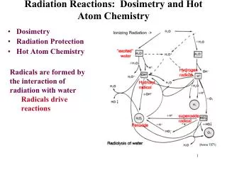
Radiation Dosimetry of the Patient. Robert L. Metzger, Ph.D. 1. Dosimetry. Radiation dosimetry is primarily of interest because radiation dose quantities serve as indicators of the risk of biologic damage to the patient

E N D
Radiation Dosimetry of the Patient Robert L. Metzger, Ph.D.
1. Dosimetry • Radiation dosimetry is primarily of interest because radiation dose quantities serve as indicators of the risk of biologic damage to the patient • The biologic effects of radiation can be classified as either deterministic (non-stochastic) or stochastic • Deterministic or non-stochastic effects are believed to be caused by cell killing • if a sufficient number of cells in an organ or tissue are killed, its function can be impaired
1. Dosimetry • Deterministic or non-stochastic effects • effects include terratogenic effects to the embryo or fetus, skin damage and cataracts • a threshold can be defined below which the effect will not occur • for doses greater than the threshold dose, the severity of the effect increases with the dose • to assess the likelihood of a deterministic effect on an organ from an imaging procedure, the dose to that organ is estimated
1. Dosimetry • A stochastic effect is caused by damage to a cell that produces genetically transformed but reproductively viable descendants • cancer and hereditary effects of radiation • probability of a stochastic effect, instead of its severity increases with dose • No dose thresholds below which the effects cannot occur • The NRC’s radiation dose limits described in Chapter 23 are intended to limit the risks of stochastic effects and to prevent the non-stochastic effects
1. Dosimetry • Entrance Skin Exposure • The radiation exposure incident on a patient is the entrance skin exposure • Skin doses are easy to measure but they are poor indicators of patient risk • They do not take into account the exposed area, penetrating power of the x-ray beam, or the radiosensitivity of the exposed region • At diagnostic energies, the f-factor (roentgen-to-rad) conversion is close to 1.0 so that dose is numerically equal to exposure
1. Dosimetry • Dose-Area Product (DAP) • Product of patient entrance skin exposure and cross-sectional area of the x-ray beam (exposed area) • Units are in mGy-cm2 or mrad-cm2 • Used in fluoroscopy
1. Dosimetry • Radiation Dose • Radiation dose is defined as the absorbed energy per unit mass but this says nothing about the total mass of tissue exposed and the distribution of the absorbed energy • Would you prefer to receive a dose of 10 mGy to the whole body or 20 mGy to the finger? • The 10 mGy whole body dose represents about 1,000 times the ionizing energy absorbed for a 70-kg person with a 35 g finger
1. Dosimetry • Imparted energy • the total amount of energy deposited in matter is called the imparted energy (Joules), is the product of the dose (Gray) and the mass (Kg) over which the energy is imparted • assume each 1-cm slice of a head CT scan delivers a 30 mGy dose to the tissue in the slice • If the scan covers 15 cm, the dose is still the same, however the imparted energy is approx. 15 times that of a single slice (you also have to consider scatter from adjacent slices, about 10-25%)
1. Dosimetry • The disadvantage of imparted energy is that it does not account for the different sensitivities of the exposed tissue to biologic damage • Effective dose is used for comparing risk of stochastic effects • E (Sv) = wT x HT • has shortcomings, wT were developed from epidemiologic data and incorporate significant uncertainties
1. Dosimetry • Organ Doses • It is possible to estimate organ doses from a given entrance skin exposure (ESE) • Organ doses are substantially lower than skin dose • For AP projections, the embryo dose will be between 1/3rd and 1/4th the ESE (in the direct beam) • For PA projections, the embryo dose will be about 1/6th of the ESE (in the direct beam) • For LAT projection, the embryo dose will be about 1/20th of the ESE (in the direct beam)
c.f. Bushberg, et al. The Essential Physics of Medical Imaging, 2nd ed., p. 59.
1. Dosimetry • Comparing ESE is useful for assessment of equipment performance and calibration, when a comprehensive analysis of effective dose is unnecessary c.f. Bushberg, et al. The Essential Physics of Medical Imaging, 2nd ed., p. 797.
1. Risk • The International Commission on Radiological Protection (ICRP) estimates the risk of fatal cancer for exposures to adults of working age to be 0.004 deaths per Sv or 0.0004 per rem • this translates to 1 cancer death per 2,500 people receiving an effective dose of 10 mSv (1 rem) • Because of the linear, no-threshold assumption used in risk estimates, risk is presumed to be proportional to the effective dose
1. Risk • Risk is proportional to the effective dose • there would be a 1 in 25,000 chance that a fatal cancer would result from an effective dose of 1 mSv (0.1 rem), or • a 1 in 500 chance of a fatal cancer from an effective dose of 50 mSv (5 rem) • The ICRP estimates the risk to be two or three times higher for infants and children and substantially lower for adults older than 50 years of age
1. Typical Absorbed and Effective doses c.f. Bushberg, et al. The Essential Physics of Medical Imaging, 2nd ed., p. 798.
1. Interventional Radiologic Procedures c.f. Bushberg, et al. The Essential Physics of Medical Imaging, 2nd ed., p. 799.
2. Radiographic Procedures Geometry for measuring the output free-in-air of a radiographic system c.f. Bushberg, et al. The Essential Physics of Medical Imaging, 2nd ed., p. 801.
2. Radiographic Procedures c.f. Bushberg, et al. The Essential Physics of Medical Imaging, 2nd ed., p. 802.
2. Radiographic Procedures Geometry for measuring the output free-in-air of a radiographic system when phototiming is used c.f. Bushberg, et al. The Essential Physics of Medical Imaging, 2nd ed., p. 804.
Question • Assuming the skin entrance dose from a single slice CT study is 5 rad, the dose for a 10 slice examination would be approximately _____ rad and the imparted energy would be ____ rad (ignore scatter). A. 5, 15 B. 15, 5 D. 50, 5 E. 5, 50
Question • The skin entrance exposure from a CT slice is 2.0 R. Ten contiguous slices are taken, then dye is injected and 10 slices are repeated. The total entrance skin exposure is about _____ R. A. 2.0 B. 2.2 D. 5.0 E. 20.0 You have to consider scatter. 25% of 2 R = 0.5. So 2.5 per scan is the rad exp. For two scans, 2.5*2 = 5.0
Question • The national average ESE for a normal 23 cm thick A/P abdomen film with a 400 speed screen-film system is: A. 13 mR B. 150 mR C. 300 mR D. 850 mR E. 3000 mR
Question • Match the exposure or dose with the appropriate item: A. 15 mR B. 40 mR C. 5 R D. 10 R E. 50 mrem 1. CT head scan ESE 2. Lateral chest ESE 3. 10 min fluoro (thin patient) 4. Monthly limit for a pregnant technologist
Question • Match the exposure or dose with the appropriate item: A. 15 mR B. 40 mR C. 5 R D. 10 R E. 50 mrem 1. CT head scan ESE – 4 to 6 R typical 2. Lateral chest ESE – 10-15 mR for PA. 2 to 3 times for Lateral 3. 10 min fluoro (thin patient) – 1-2 R/min for thin patient 4. Monthly limit for a pregnant technologist – 0.5 mSv or 50 mrem