Download
1 / 1
10 likes | 183 Vues
Developmental Psychobiology Masoud Asiaei 1 Jalal Solati 1 Ali-Akbar Salari 2 1 Department of Biology Faculty of Science, Karaj Branch Islamic Azad University P.O. Box 31485-313, Karaj, Iran E-mail: solati@kiau.ac.ir 2 Department of Biology Islamic Azad University

E N D
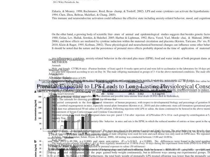
Developmental Psychobiology Masoud Asiaei1 Jalal Solati1 Ali-Akbar Salari2 1Department of Biology Faculty of Science, Karaj Branch Islamic Azad University P.O. Box 31485-313, Karaj, Iran E-mail: solati@kiau.ac.ir 2Department of Biology Islamic Azad University North Tehran Branch, Tehran, Iran Prenatal Exposure to LPS Leads to Long-Lasting Physiological Consequences in Male Offspring ABSTRACT: Growing evidence suggests that early life events are critical determinants for disorders later in life. According to a comprehensive number of epidemiological/animal studies, exposure to lipopolysaccharide, causes alteration in pro-inflammatory cytokine levels, hypothalamic–pituitary–adrenal functioning and the hormonal system which may contribute to behavioral and neurological injuries. In this study we investigated the effects of lipopolysaccharide adminis- tration on physiological parameters in pregnant dams and their male offspring aged 9 weeks. In gestational Day 10, pregnant mice were injected intraprito- neally with Salmonella enterica lipopolysaccharide to model prenatal exposure to infection. The following results were obtained for offspring from dams stressed during pregnancy: (a) reduced anxiety-related behavior in the elevated plus maze; (b) reduced food and water intake; (c) reduced body weight from birth up to postnatal Day 40. The observed data provide experimental evidence showing that prenatal stress can have complex and long-lasting physiological/ behavioral consequences in offspring. 2011 Wiley Periodicals, Inc. Dev Psychobiol 53: 828–838, 2011. Keywords: lipopolysaccharide; prenatal stress; pro-inflammatory cytokine; corti- costerone; body weight; food intake; water intake; C57BL/6 mice INTRODUCTION Lipopolysaccharide (LPS), an endotoxin produced from the cell walls of gram-negative bacteria, acts as a non- specific immunostimulant to induce a severe inflamma- tory response by initiating multiple intracellular signaling events, including the activation of nuclear factor kb (NF-kb), which ultimately leads to the syn- thesis and release of cytokines, such as pro-inflamma- tory cytokine tumor necrosis factor alpha (TNF-a), interleukin 1b (IL-1b) and interleukin 6 (IL-6) from macrophages (Anisman, Hayley, Turrin, & Merali, 2002; Anisman, Kokkinidis, & Merali, 2002; Anisman, Received 8 December 2010; Accepted 25 April 2011 Correspondence to: J. Solati Published online 31 May 2011 in Wiley Online Library (wileyonlinelibrary.com). DOI 10.1002/dev.20568 2011 Wiley Periodicals, Inc. Zaharia, & Meaney, 1998; Bachmanov, Reed, Beau- champ, & Tordoff, 2002). LPS and some cytokines can activate the hypothalamic–pituitary–adrenal (HPA) axis which leads to increase in plasma concentrations of adrenocorticotropic hormone (ACTH), glucocorticoids and also corticotrophin releasing factor (CRF) (Ban et al., 1993; Barker et al., 1993; Becskei et al., 2008; Bell & Hallenbeck, 2002), causing changes in the brain neurochemistry (Betancur, Lledo, Borrell, & Guaza, 1994; Chen, Zhou, Beltran, Malellari, & Chang, 2005). This immune and neuroendocrine activation could influence the effective state including anxiety-related behavior, mood, and cognition that have clinical impor- tance (Degroot, Kashluba, & Treit, 2001; Delrue, Dele- planque, Rougepont, Vitiello, & Neveu, 1994). Animal and human studies showed that there is an increase of above-mentioned cytokines both in systemic and mRNA levels, especially in the brain of rodents follow- ing peripheral exposure to LPS (Dunn, 1988, 1989; Dunn & Berridge, 1990). On the other hand, a growing body of scientific liter- ature of animal and epidemiological studies suggests that besides genetic factors, environmental factors, like maternal stress can have long-lasting effects on physical development, neurochemistry, behavior and immunocompetence of the offspring, hence this phenomenon has been denoted as ‘‘fetal programming’’ (Barker et al., 1993; File, 1996, 2001). According to several scientific literatures, these maternal stresses are very important elements in the provocation or exacer- bation of a wide range of physiological/behavioral and psychological/mental disturbances (Fride & Weinstock, 1988; Golan, Lev, Hallak, Sorokin, & Huleihel, 2005; Harbuz & Lightman, 1992; Hava, Vered, Yael, Morde- chai, & Mahoud, 2006). Maternal stress can lead to elevated levels of maternal stress hormones, notably, it is well established that HPA hormones play a critical role in the stress response (Johnson, Kamilaris, Chrou- sos, & Gold, 1992). In rodents and non-human primate species, it was found that prenatal stress, including exposure to endotoxin, can alter HPA axis and brain neurotransmitter systems in the offspring (Kapoor, Dunn, Kostaki, Andrews, & Matthews, 2006; Karrow, 2006), and these effects are mediated by cytokine induction within the maternal circulation and placenta (Kirsten, Taricano, Flo´ rio, Palermo-Neto, & Bernardi, 2010; Klein & Rager, 1995; Kofman, 2002). These physiological and neurochemical/hormonal changes can influence some other behaviors like eating and drink- ing. Food and water intakes correlated positively, and this may be due to their mutual dependence on body size, but an additional mechanism directly linking food and water intakes may also be involved (Bachmanov et al., 2002; Kraly, 1984). It should be noted that the nature and the persistence of prenatal stress effects probably depend on the time of application of maternal stress relative to the fetal stage of development (Merlot, Couret, & Otten, 2008). There is emerging evidence suggesting that inflamma- tory events associated with immunological events in early/middle fetal life (e.g., GD 8-10 in rats and mice) are likely to have more stronger neurodevelopmental impacts than late-pregnancy inflammations. These maternal immune activations during early/middle preg- nancy impede with cell proliferation, differentiation, migration, target selection, and synapse maturation, finally leading to several brain and behavioral abnor- malities in adulthood (Kirsten et al., 2010). The present study proceeded to investigate some of the neuroendo- crine and behavioral consequences of prenatal exposure to LPS on both pregnant C57BL/6 mice in gestational day (GD) 10 and their male offspring aged 9 weeks. Therefore, we assessed the effect of prenatally adminis- tered LPS on the concentration of corticosterone and pro-inflammatory cytokines, anxiety-related behavior in the elevated plus maze (EPM), food and water intake of both pregnant dams and their male offspring at adulthood and also the body weight of offspring from birth to postnatal day (PND) 40. METHODS Animals Male and female C57BL/6 mice (Pasteur Institute of Iran) aged 6–8 weeks upon arrival and were left to acclimatize to the laboratory for 10 days prior to testing. Mice were main- tained in groups of 5 in standard polypropylene cages, to avoid behavioral changes that may result from single housing which can usually be an increase in aggressive and fear-like behavior. The animals were allowed free access to food and water at all times and were maintained on a 12 hr light/dark schedule (lights on 07:00 hr) in a controlled temperature (23 18C). For mating purposes, three females were housed overnight with two males starting at 19:00 hr. Each female mouse was visually inspected for the presence of a vaginal plug the next morning at 07:00 hr. The presence of plug was designed as day 0 of gestation. The pregnant mice (N ¼ 80) were divided randomly into four groups. Forty of these mice (N ¼ 10 in each group) were scarified for measuring cyto- kines and corticosterone and others were left to labor (N ¼ 10 in each group). Following delivery, litters remained intact to avoid confounding changes in maternal behavior. The litters remained with their mothers until weaning (Day 21) and were separated according to sex on Day 36. The male offspring maintained in groups of 3–4 in the above-mentioned conditions. The male offspring were distributed into control and experimental groups (N ¼ 10/group; two pups were selected from each mother for the next experiments). The study was approved by the Ethics Committee of Karaj Islamic Azad University and experimental protocol is in compliance with the National Institutes of Health Guide for Care and Use of Laboratory Animals (Publication No. 85-23, revised 1985). Prenatal Administration of LPS Lipopolysaccharide (Salmonella enterica serotype entritidis, L6011, Sigma Aldrich, Saint Louis, MO) was dissolved in ster- ile pyrogen-free saline before use. The dams were randomly assigned to a saline control group and LPS groups. The dams in the LPS groups were administered a single intraperitoneal (i.p.) injection of 50, 100, or 150 mg/kg LPS on day 10 of pregnancy. The dams in the control group were administered a single i.p. injection of saline on day 10 of pregnancy. It is important to note that although maternal exposure to LPS causes some disturbances, it can vary depending on doses of LPS, potency of LPS, age, sex, and interactions with environ- mental and genetic risk factors (Bell & Hallenbeck, 2002; Urakubo, Jarskog, Lieberman, & Gilmore, 2001). The dosage of LPS we chose could induce systemic inflammation, resulting in a low percentage of fetal anomalies, but not abortion and possible intra-uterine fetal death (IUFD) (Bell & Hallenbeck, 2002). The rationale for choosing gestational day 10 was that this period corresponds to the first-to-second trimesters of human pregnancy, with respect to developmental biology and percentage of gestation from mice to human (Kaufman, 1992). Other scientific literatures state that this time phase is the period of early fetal brain development (Wei, Li, & Zhou, 2007), cerebral organogenesis in mice, especially neural-plate formation (Kirsten et al., 2010) and also embryonic stem cell formation (gestational period 0.38–0.53 in rodents) which is one of the main periods of vulnerability of the immune system to environmental insults (Merlot et al., 2008). In addition, other investigators suggest that maternal infection from early to mid pregnancy is more likely to be related to long-lasting develop- mental brain and behavioral abnormalities in the offspring (Mednick, Machon, Huttunen, & Bonett, 1988; Rodier & Hyman, 1998). Each dam was administered 50 ml saline or LPS solution. Following injection with LPS or saline, the dams continued to be housed in the above-mentioned conditions. As we know, LPS can disrupt the blood–brain barrier (BBB) if adminis- tered topically to the cerebral microcirculation or intracister- nally, and it is possible that high doses of LPS cross the placenta into the fetal circulation (Urakubo et al., 2001). Low doses of LPS were selected in order to prevent possible direct exposure of fetus to LPS. With this dose and route of application we observed no abortion in our endotoxin groups. All injections were given between 13:00 and 14:00 hr. Measurement of Serum Corticosterone and Cytokine Concentration Maternal serum from trunk blood of pregnant dams was pre- pared 1.5 hr after injection of LPS/saline (N ¼ 10 in each group) by centrifugation at 5,000g for 12 min, aliquoted and then stored at 808C until the cytokine/corticosterone assays were performed. Concentrations of corticosterone (KT-510, Kamiya Biomedical Company, Seattle, WA), IL-1b (IB49700, Immuno-Biological Laboratories, Minneapolis, MN), IL-6 (BioSource International, CA), and TNF-a (CT 302, Ucytech, Utrecht, The Netherlands) were determined by using commer- cial ELISA kits according to the manufacturer’s protocol. Trunk blood was collected for preparation of blood serum of male offspring in their 9th week (PND 62) and measurement of serum corticosterone and cytokine concentration was carried out again in the same way. All samples and standards were assayed in duplicate. The pregnant mice and their male offspring were selected randomly. Anxiety Test One of the most popular tests of anxiety-like behavior in mice and rats is the EPM, in which the reduced number of entries or time spent in the open arms of the EPM suggests the operation of anxiety-like processes. This wooden, plus- shaped apparatus was elevated to the height of 50 cm above the floor and consists of two open arms (30 cm 5 cm), two enclosed arms (30 cm 5 cm 15 cm), and central platform (5 cm 5 cm) each with an open roof. The maze was placed in the center of a quiet and dimly lit room. The mice behavior was directly observed using a mirror, sus- pended at an angle above the maze. Behavioral data were collected by a ‘‘blind’’ observer who quietly sat 1 m behind one of the closed arms of the maze, using a chronometer. The anxiety test of the offspring was carried out at postnatal Day 61. It was repeated three times, three separate cohorts of male offspring were used for tests and each mouse was only used on EPM once. We repeated this test three times due to our different results on EPM from other investigations and the results represent an average of the three test sessions. For test- ing purpose, male offspring were placed individually in the center portion of the plus-maze, facing one of the open arms. The observer measured: (1) the time spent in the open arms, (2) the time spent in the closed arms, (3) the number of entries into the open arms, and (4) the number of entries into the closed arms during the 5-min test period. An entry was defined as all four paws in the arm. The elevated plus-maze was thoroughly cleaned with distilled water following the testing of each animal to avoid possible biasing effects due to odor clues left by previous mice. For the purpose of analysis, open-arm activity was quantified as the amount of time that the mice spent in the open arms (OAT) relative to the total amount of time spent in any arm (open/total 100), and the number of entries into the open arms (OAE) was quantified relative to the total number of entries into any arm (open/ total 100). The total number of open arms entered, as well as the total number of closed arms entered were used as indexes of general locomotor activity (LMA) (Solati, Zarrindast, & Salari, 2010; Zarrindast, Solati, Oryan, & Parivar, 2008). All testing was conducted between 13:00 and 16:00 hr. Body Weight The body weights of male offspring ( 0.1 g) were regularly monitored at 13:00 hr every 10 days during the experiment from birth (PND 0) to PND 40. Food and Water Intake In order to find out whether i.p. exposure to LPS during preg- nancy affects food/water intake in dams or in their male off- spring under our experimental conditions, a feeding/drinking study was conducted. Mice had free access to food and water before the experiments began. Each cage contained 3 or 4 mice which were given with the same amount of food and water. Their food intake was measured the following day by subtracting the uneaten food manually. The amount of water ingested in our experiment was measured with 0.2 ml gradu- ated glass burettes adapted with a metal drinking spout. Immediately after injection of LPS/saline in pregnant dams, each mouse was returned to its cage and we measured the cumulative water intake manually the following day. These measurements were done in 3 days after injection of the LPS/ saline in respect to pregnant dams (N ¼ 10 in each group) and in 5 days in PND 56-60 in male offspring (N ¼ 10 in each group) and it was calculated as food (in grams 0.1 g)/ water (in ml 0.2 ml) intake per mice per day. Measurements were performed by the same experimenter and we used separ- ate cohorts of pregnant dams for food/water intake, but the same groups of male offspring were used for food/water intake and anxiety test on EPM. Statistical Analysis Data were analyzed using SPSS. Since data displayed normal distribution and homogeneity of variance, one-way ANOVA was used for comparison between the effects of different doses of LPS with vehicle. Differences with p < 0.05 between experimental groups at each point were considered statistically significant. RESULTS Effects of LPS on Serum Concentrations of TNF-a, IL-1b, and IL-6 in Pregnant Mice and Their Male Offspring The effects of LPS treatment on TNF-a, IL-1b, and IL-6 in maternal serum were analyzed. In response to LPS challenge, TNF-a (F3,36 ¼ 161.825, p < 0.001), IL-1b (F3,36 ¼ 39.468, p < 0.000) and IL-6 (F3,36 ¼ 9.968, p < 0.001) in maternal serum were increased 1.5 hr after LPS administration (except for IL-6 in 50 mg/kg LPS; Fig. 1). Table 1 shows that there were no signifi- cant differences between prenatally LPS and saline- treated offspring in serum concentrations of TNF-a (F3,36 ¼ 1.971, p < 0.145), IL-1b (F3,36 ¼ 1.403, p < 0.266) and IL-6 (F3,36 ¼ 1.496, p < 0.241) in their 9th week (PND 62). Effects of LPS on Serum Concentrations of Corticosterone in Pregnant Mice and Their Male Offspring The effects of LPS treatment on serum corticosterone level of pregnant dams and their male offspring are shown in Figure 1 and Table 1. One way ANOVA confirmed that in pregnant dams, high doses of LPS (100 or 150 mg/kg) increased serum corticosterone levels relative to saline-treated animals (F3,36 ¼ 8.937, p < 0.002) (Fig. 1). But prenatal LPS exposure does not have any significant effect on serum concentrations of corticosterone in male offspring in their 9th week (PND 62) (F3,36 ¼ 3.442, p < 0.052) (Tab. 1). Effects of LPS on Anxiety-Related Behaviors in Male Offspring Figure 2 shows data on the effects of prenatal exposure to LPS on anxiety-related behavior of male offspring in the EPM. One-way ANOVA revealed that prenatal exposure to LPS has increased the percentage of open FIGURE 1 Effect of intraperitoneal injection of saline (50 ml/mouse) or LPS (50, 100, or 150 mg/kg) on serum level of TNF-a, IL-1b, IL-6 (a) and corticosterone (b) in pregnant dams. Each bar is mean SEM. N ¼ 10. p < 0.05, p < 0.01, and p < 0.001, when compared to the saline treated group. arm time (F3,36 ¼ 49.396, p < 0.000) and open arm entries (F3,36 ¼ 8.843, p < 0.000). No differences were found in the total number of arm entries in the plus-maze (open-arms þ closed-arms) (F3,36 ¼ 1.604, p < 0.215). Considering all of these effects, the pres- ently observed behavioral data suggest low levels of anxiety in prenatally LPS-treated offspring which was not reported like this before. Effects of Prenatal Exposure to LPS on Body Weight in Male Offspring During postnatal development, all the pups gained body weight continuously, but in different ways among our experimental groups. The birth weight (PND 0) of prenatally LPS-treated offspring (LPS 50: 1.31 0.33 g; LPS 100: 1.20 0.33 g; LPS 150: 1.20 0.32 g) was lower than prenatally saline-treated offspring (1.62 0.31 g) (F3,36 ¼ 36.282, p < 0.000). Furthermore, the total body weight of prenatally LPS-treated offspring was lower than the prenatally saline-treated offspring during 40 days of age [PND 10 (F3,36 ¼ 26.399, Table 1. Effect of Prenatal Exposure to LPS on Serum Level of Pro-Inflammatory Cytokines and Corticosterone in Male Offspring in PND 62 (Mean SEM, N ¼ 10 in Each Group) p < 0.000), PND 20 (F3,36 ¼ 25.849, p < 0.000), PND 30 (F3,36 ¼ 8.174, p < 0.001), PND 40 (F3,36 ¼ 5.366, p < 0.006)] (Fig. 3). It is important to note that we observed no miscarriage or fetal loss among our groups during experiment. Effects of LPS on Food and Water Intake in Pregnant Mice and Their Male Offspring As can be clearly seen in Figure 4, i.p. administration of LPS reduced food intake in the first day after injec- tion in pregnant dams (F3,36 ¼ 13.465, p < 0.000). During the 2nd (F3,36 ¼ 2.375, p < 0.095) and 3rd (F3,36 ¼ 1.685, p < 0.197) day after challenge, there were no significant differences between LPS and saline-treated dams in food intake. In other words, feeding then returned to normal over the next 48–72 hr as the LPS effect dissipated. It is also important to note that there were significant decreases in food intake FIGURE 2 Effects of prenatal exposure to LPS on anxiety- related behavior in offspring in the elevated plus-maze. Male offspring prenatally exposed to saline (50 ml/mouse) or LPS (50, 100, or 150 mg/kg) and anxiety test of these offspring carried out at postnatal Day 61. Each bar is mean SEM. Open arm time (OAT), open arm entries (OAE) or locomotor activity (LMA). N ¼ 10. p < 0.05, p < 0.01, and p < 0.001 when compared to the saline treated group. between prenatally LPS or saline-treated male offspring at postnatal Day 56–60 [PND 56 (F3,36 ¼ 37.029, p < 0.000), PND 57 (F3,36 ¼ 20.638, p < 0.000), PND 58 (F3,36 ¼ 30.582, p < 0.000), PND 59 (F3,36 ¼ 45.155, p < 0.000), PND 60 (F3,36 ¼ 36.505, p < 0.000)] (Fig. 4). The intraperitoneal injection of small amounts of endotoxin taken from Salmonella enterica significantly decreased and almost completely interrupted the water intake of pregnant dams. One day after LPS exposure there was a significant decrease in water intake among all dams (F3,36 ¼ 47.734, p < 0.000). In other words, in the experiment presented in Figure 5, 50 mg/kg of endotoxin was sufficient to alter water intake. There were no significant differences in the water intake of dams which received the smallest dose of endotoxin, compared to the control group, but the inhibition per- sisted longer as the dose was increased (F3,36 ¼ 20.714, p < 0.000). With 100 or 150 mg/kg of endo- toxin, the daily water intake in pregnant dams did not return to normal until the end of the 3rd day after injec- tion (F3,36 ¼ 4.975, p < 0.008), (see Fig. 5). The water FIGURE 3 Effect of prenatal exposure to saline (50 ml/ mouse) or LPS (50, 100, or 150 mg/kg) on body weight of male offspring from birth day to PND 40. The endotoxin offspring had a lower body weight from birth day up to PND 40. Each bar is mean SEM. N ¼ 10. p < 0.01 and p < 0.001 when compared to the saline treated group. FIGURE 4 Effect of intraperitoneal injection or prenatal exposure to saline (50 ml/mouse) or LPS (50, 100, or 150 mg/kg) on food intake in pregnant dams and their male offspring. Endotoxin dams ate less than saline animals just in first day after LPS injection, while endo- toxin offspring ate less than saline offspring during experiment. Each bar is mean SEM. N ¼ 10. p < 0.001 when compared to the saline treated group. intake in prenatally LSP-treated male offspring was lower compared to prenatally-saline treated offspring in PND 56–60 [PND 56 (F3,36 ¼ 78.004, p < 0.000), PND 57 (F3,36 ¼ 109.789, p < 0.000), PND 58 (F3,36 ¼ 137.651, p < 0.000), PND 59 (F3,36 ¼ 161.790, p < 0.000), PND 60 (F3,36 ¼ 6.552, p < 0.002)] (Fig. 5). Different studies showed that the inhibition of water intake was essentially the same, despite the strain of Gram-negative species from which the endotoxin was prepared. FIGURE 5 Effect of intraperitoneal injection or prenatal exposure to saline (50 ml/mouse) or LPS (50, 100, or 150 mg/kg) on water intake in pregnant dams and their male offspring. Each bar is mean SEM. N ¼ 10. p < 0.05, p < 0.01, and p < 0.001, when com- pared to the saline treated group. DISCUSSION There is a consensus that severe environmental or psychological stress, including exposure to LPS, results in the release of glucocorticoids, pro-inflammatory cytokines, ACTH and CRF in maternal blood stream. These changes could operate directly or indirectly by regulating the activation of HPA axis and changes in placental metabolism on fetal development. Among these candidates, maternal glucocorticoids and pro- inflammatory cytokines are the main candidates for the mediation of prenatal stress to the fetus (Harbuz & Lightman, 1992; Johnson et al., 1992; Weinstock, Pol- tyrev, Schorer-Apelbaum, Men, & McCarty, 1998). Glucocorticoids are known for their central roles in immunocompetence in anxiety circumstances (Dunn, 1989; Dunn & Berridge, 1990; Merlot et al., 2008; Weinstock et al., 1998) and they can apply many organ- izational effects on prenatal tissue development, includ- ing HPA development, leading to changes in the HPA axis function that persist throughout life (Merlot et al., 2008; Muglia, Jacobson, Dikkes, & Majzoub, 1995). In this study, we measured the level of corticosterone, major murine glucocorticoid hormone, and pro-inflam- matory cytokines in both pregnant dams and their male offspring. As described in the results, we observed an increased level of pro-inflammatory cytokines (except for IL-6 in 50 mg/kg LPS) and corticosterone in preg- nant dams which received 100 or 150 mg/kg LPS, but not in their male offspring. These elevations of maternal cytokines and glucocorticoids can result in growth retardation and behavioral alterations in the off- spring (Anisman, Hayley, et al., 2002; Anisman, Kokki- nidis, et al., 2002). The possible mechanism is that prenatal stress such as exposure to high level of gluco- corticoids has long-term consequences on child, includ- ing on neurodevelopment (Talge, Neal, & Glover, 2007; Van den Bergh, Mulder, Mennes, & Glover, 2005). Glucocorticoids inhibit hippocampal cell pro- liferation (Gould, Cameron, Daniels, Woolley, & McE- wen, 1992; Reul et al., 1994) and exert their action via direct influence on ontogeny of fetal cells by reaching the fetal organs. In this respect human studies show that increased cortisol exposure affects the expression of over a thousand genes in fetal brain cells and this exposure affect the function of the placenta, including the expression and activity of 11beta-hydroxysteroid dehydrogenase 2 (11b-HSD2), the main barrier to the placental passage of glucocorticoids (Glover, Bergman, Sarkar, & O’Connor, 2009; Mairesse et al., 2007; Salaria et al., 2006; Welberg, Thrivikraman, & Plotsky, 2005). In humans lower placental levels of 11b-HSD2 have been found to be associated with intrauterine growth restriction (Dy, Guan, Sampath-Kumar, Richardson, & Yang, 2008; Glover et al., 2009). Other human studies show that synthetic glucocorticoids that cross the placenta can affect infant neurodevelopment (Sizonenko et al., 2006). On the other hand, animal studies also demonstrate long-term effects of prenatal administration of synthetic glucocorticoids, such as dexamethasone, on offspring brain development and behavior. For example, some animal works show that prenatal exposure to raised glucocorticoids can damage the brain and caused a reduction in hippocampal vol- ume in rodents and monkeys (Bergman, Sarkar, Glover, & O’Connor, 2010; Mitra & Sapolsky, 2008; Uno et al., 1994). Thus, maternal hormones seem to be good candidates for communication between the dam and developing fetus (Joffe, 1969; Merlot et al., 2008). Although direct evidence for these causal links is lack- ing, and the mechanisms by which infection-induced elevation of cytokine and glucocorticoid levels during fetal life can interfere with neurodevelopmental events remain poorly understood. It has been suggested that infections and inflamma- tory processes may be causative factors in emotional disorders, including anxiety (Belzung & Griebel, 2001; File, 2001; Hogg, 1996). To explore this hypothesis, we measured the anxiety-related behavior of male off- spring on EPM. Prenatal LPS exposure without loco- motor impairment in the elevated plus maze, increased the percentage of open arm times and open arm entries in adult male offspring, indicating the induction of anxiolytic response by prenatal immune challenge, which do not agree with others’ obtained in similar contexts (Dunn & Berridge, 1990; Fride & Weinstock, 1988, 1989). Though, at first these results appear con- tradictory, the discrepancy may be related to the g-ami- nobutyric acid (GABA) system. Prenatal stress has been found to affect the GABA system in different brain regions, however, different stress paradigms have different effects on the GABA system (Bowers, Cullinan, & Herman, 1998; Gruen, Wenberg, Elahi, & Friedhoff, 1995; Montpied et al., 1993). GABA, which is the main inhibitory neurotransmitter of brain, has a well known and very important role in the modulation of anxiety. In addition, it is well known that GABA- ergic agents and drugs have strong anxiolytic effects in humans and animals. One feasible mechanism is that prenatal exposure to endotoxin changes the sensitivity of GABAA receptor and finally modifies the behavioral responses to stress in adulthood (Jaiswal & Bhatta- charya, 1993; Kellogg, Taylor, Rodriguez-Zafra, & Pleger, 1993). In fact, LPS may have negated the effects of stress on neonate mice, for example, through suppression of a stress-induced surge of corticosterone and altering the composition and function of the GABAA receptor complex by means of a transcriptional mechanism (Stone et al., 2001). Stone et al. (2001) demonstrated that elevation in the level of glucocorti- coids can affect the GABAergic system and change the expression of GABAA receptor subunit mRNA levels in the brain regions that are involved in modulation anxiety, like hippocampus. This elevation also affects mRNAs for glutamic acid decarboxylase (GAD) iso- forms, the enzyme that converts glutamate to GABA. Prenatal increase of corticosterone level increases GAD67 mRNA in hippocampus. Increased inhibitory input by local GABA neurons may discuss the effects of prenatal stresses on reduced anxiety-related behav- iors in adulthood. Therefore, there is a possibility that prenatal activation of HPA axis by LPS may increase the activity of GABAergic system in the brain regions like hippocampus and in this way decrease the anxiety level of offspring (Stone et al., 2001). In sum, it is clear that our results cannot directly prove this phenomenon and surely need to be replicated before firm conclusions can be drawn. In other parts of our study we examined the effects of LPS exposure on ingestive behaviors of pregnant dams and their male offspring. In this part of the study we observed the reduction of food and water intake in both pregnant dams and their male offspring (PND 56–60). The central mechanisms underlying anorexia during exposure to bacterial components are not fully understood. Several studies confirmed that all systems, signals, and switches (physiologic, metabolic, neuro- logic, anatomic, endocrine, behavioral, etc.) can influ- ence food and water intake (Bachmanov et al., 2002; Kraly, 1984) and Moyer, Herrenkohl, and Jacobowitz (1978) suggested that prenatal stress may cause perma- nent changes in noradrenaline hypothalamic content with a possible anorexic effect (Moyer et al., 1978). Our results, which concur with other reports in the literature (Maccari et al., 2003; Maccari & Morley- Fletcher, 2007; Seckl, 2001; Tamashiro, Terrillion, Hyun, Koenig, & Moran, 2009), demonstrated that exposure to increased levels of corticosterone and pro- inflammatory cytokines during prenatal stage, induce adverse effects on ingestive behaviors in later life. These results thus highlight the long-term influences of prenatal exposure to LPS (which leads to maternal malnutrition, anxiety and hormonal imbalance during pregnancy) on ingestive behaviors in mice. Although it needs replication in larger cohorts before firm con- clusions can be drawn. Previous reports on the effects of prenatal stresses on body weight are contradictory. Some authors reported that prenatal stress leads to reduced body weight of pups (Armario, Restrepo, Castellanos, & Balasch, 1985; Klein & Rager, 1995; Patin et al., 2002; Pollard, 1984; Salgado, Martinez, & Tarres, 1977; Valle´e, Mayo, Maccari, Le Moal, & Simon, 1996), whereas other studies failed to find an effect (Fride, Dan, Feldon, Halevy, & Weinstock, 1986; Glo¨ ckner & Karge, 1991; Hughes & Beveridge, 1990). The discrep- ancies between studies could be due to differences in the prenatal stress protocols employed. Our study indicates that prenatal exposure to Salmonella endo- toxin decreases fetal body weight at term, indicating a fetal growth restriction. In addition, the male offspring with prenatal exposure to LPS showed decrease in body weights when they grew up, in which the duration of the effects was directly related to the dose injected. The possible mechanism(s) is that decrease in body weight of the offspring might have been induced by long-term effects of maternal nutritional deficiency, hyperactivity of the HPA axis (Levitt et al., 2000; Phil- lips et al., 1998; Reynolds & Phillips, 2000), excessive glucocorticoid level in fetal environment (Krieg et al., 1992) and/or alteration in organ maturation (Novy & Walsh, 1983; Reinisch, Simon, Karow, & Gandelman, 1978). Although the mechanisms underlying these phenomena are still unclear, further understanding could be gained by investigating the consequences of prenatal manipulations. CONCLUSION In summary, this study demonstrated that exposure to endotoxin during fetal life can lead to long-lasting physiological consequences most of them detrimental for the animal during adulthood. This conclusion was based on the following data observed in dams exposed to LPS in GD10 during pregnancy and their male off- spring versus the control group: (1) Increased pro- inflammatory cytokine and corticosterone levels in maternal serum, (2) Decrease in anxiety-related behav- ior in male offspring, (3) Decrease in food and water intake in pregnant dams, and interestingly in their male offspring, (4) Restriction in the body weight of male offspring up to PND 40. REFERENCES Anisman, H., Hayley, S., Turrin, N., & Merali, Z. (2002). Cytokines as a stressor: Implications for depressive illness. The International Journal of Neuropsychopharmacology, 5(04), 357–373. Anisman, H., Kokkinidis, L., & Merali, Z. (2002). Further evidence for the depressive effects of cytokines: Anhedo- nia and neurochemical changes. Brain, Behavior, and Immunity, 16(5), 544–556. Anisman, H., Zaharia, M., & Meaney, M. (1998). Do early- life events permanently alter behavioral and hormonal responses to stressors? International Journal of Develop- mental Neuroscience, 16(3–4), 149–164. Armario, A., Restrepo, C., Castellanos, J., & Balasch, J. (1985). Dissociation between adrenocorticotropin and corticosterone responses to restraint after previous chronic exposure to stress. Life Sciences, 36(22), 2085–2092. Bachmanov, A., Reed, D., Beauchamp, G., & Tordoff, M. (2002). Food intake, water intake, and drinking spout side preference of 28 mouse strains. Behavior Genetics, 32(6), 435–443. Ban, E., Marquette, C., Sarrieau, A., Fitzpatrick, F., Fillion, G., Milon, G., et al. (1993). Regulation of lnterleukin-1 receptor expression in mouse brain and pituitary by lipo- polysaccharide and glucocorticoids. Neuroendocrinology, 58(5), 581–587. Barker, D., Godfrey, K., Gluckman, P., Harding, J., Owens, J., & Robinson, J. (1993). Fetal nutrition and cardiovascular disease in adult life. The Lancet, 341(8850), 938–941. Becskei, C., Riediger, T., Herna´dfalvy, N., Arsenijevic, D., Lutz, T., & Langhans, W. (2008). Inhibitory effects of lip- opolysaccharide on hypothalamic nuclei implicated in the control of food intake. Brain, Behavior, and Immunity, 22(1), 56–64. Bell, M., & Hallenbeck, J. (2002). Effects of intrauterine inflammation on developing rat brain. Journal of Neuro- science Research, 70(4), 570–579. Belzung, C., & Griebel, G. (2001). Measuring normal and pathological anxiety-like behaviour in mice: A review. Behavioural Brain Research, 125(1–2), 141–149. Bergman, K., Sarkar, P., Glover, V., & O’Connor, T. G. (2010). Maternal prenatal cortisol and infant cognitive development: Moderation by infant-mother attachment. Biological Psychiatry, 67(11), 1026–1032. Betancur, C., Lledo, A., Borrell, J., & Guaza, C. (1994). Cor- ticosteroid regulation of IL-1 receptors in the mouse hippocampus: Effects of glucocorticoid treatment, stress, and adrenalectomy. Neuroendocrinology, 59(2), 120–128. Bowers, G., Cullinan, W. E., & Herman, J. P. (1998). Region- specific regulation of glutamic acid decarboxylase (GAD) mRNA expression in central stress circuits. Journal of Neuroscience, 18(15), 5938. Chen, R., Zhou, H., Beltran, J., Malellari, L., & Chang, S. (2005). Differential expression of cytokines in the brain and serum during endotoxin tolerance. Journal of Neuro- immunology, 163(1–2), 53. Degroot, A., Kashluba, S., & Treit, D. (2001). Septal GABA- ergic and hippocampal cholinergic systems modulate anxiety in the plus-maze and shock-probe tests. Pharma- cology Biochemistry and Behavior, 69(3–4), 391–3399. Delrue, C., Deleplanque, B., Rougepont, F., Vitiello, S., & Neveu, P. (1994). Brain monoaminergic, neuroendocrine, and immune responses to an immune challenge in relation to brain and behavioral lateralization. Brain, Behavior, and Immunity, 8(2), 137–152. Dunn, A. (1988). Systematic interleukin-1 administration stimulates hypothalamic norepinephrine metabolism parallelling the increased plasma corticosterone. Life Sci- ences, 43(5), 429–435. Dunn, A. (1989). Psychoneuroimmunology for the psycho- neuroendocrinologist: A review of animal studies of nervous system-immune system interactions. Psychoneur- oendocrinology, 14(4), 251–274. Dunn, A., & Berridge, C. (1990). Physiological and behavior- al responses to corticotropin-releasing factor adminis- tration: Is CRF a mediator of anxiety or stress responses. Brain Research Reviews, 15(71), 100. Dy, J., Guan, H., Sampath-Kumar, R., Richardson, B., & Yang, K. (2008). Placental 11 [beta]-hydroxysteroid dehydrogenase type 2 is reduced in pregnancies compli- cated with idiopathic intrauterine growth restriction: Evidence that this is associated with an attenuated ratio of cortisone to cortisol in the umbilical artery. Placenta, 29(2), 193–200. File, S. (1996). Recent developments in anxiety, stress, and depression. Pharmacology Biochemistry and Behavior, 54(1), 3–12. File, S. (2001). Factors controlling measures of anxiety and responses to novelty in the mouse. Behavioural Brain Research, 125(1–2), 151–157. Fride, E., Dan, Y., Feldon, J., Halevy, G., & Weinstock, M. (1986). Effects of prenatal stress on vulnerability to stress in prepubertal and adult rats. Physiology & Behavior, 37(5), 681–687. Fride, E., & Weinstock, M. (1988). Prenatal stress increase anxiety related behavior and alters cerebral lateralization of dopamine activity. Life Sciences, 42(10), 1059–1065. Fride, E., & Weinstock, M. (1989). Alterations in behavioral and striatal dopamine asymmetries induced by prenatal stress. Pharmacology Biochemistry and Behavior, 32(2), 425–430. Glo¨ ckner, R., & Karge, E. (1991). Influence of chronic stress before and/or during gestation on pregnancy outcome of young and old Uje: WIST rats. Journal of Experimental Animal Science, 34(3), 93. Glover, V., Bergman, K., Sarkar, P., & O’Connor, T. G. (2009). Association between maternal and amniotic fluid cortisol is moderated by maternal anxiety. Psychoneuroen- docrinology, 34(3), 430–435. Golan, H., Lev, V., Hallak, M., Sorokin, Y., & Huleihel, M. (2005). Specific neurodevelopmental damage in mice off- spring following maternal inflammation during pregnancy. Neuropharmacology, 48(6), 903–917. Gould, E., Cameron, H. A., Daniels, D. C., Woolley, C. S., & McEwen, B. S. (1992). Adrenal hormones suppress cell division in the adult rat dentate gyrus. Journal of Neuro- science, 12(9), 3642. Gruen, R. J., Wenberg, K., Elahi, R., & Friedhoff, A. J. (1995). Alterations in GABAA receptor binding in the pre- frontal cortex following exposure to chronic stress. Brain Research, 684(1), 112–114. Harbuz, M., & Lightman, S. (1992). Stress and the hypothalamo-pituitary-adrenal axis: Acute, chronic and immunological activation. Journal of Endocrinology, 134(3), 327. Hava, G., Vered, L., Yael, M., Mordechai, H., & Mahoud, H. (2006). Alterations in behavior in adult offspring mice fol- lowing maternal inflammation during pregnancy. Develop- mental Psychobiology, 48(2), 162–168. Hogg, S. (1996). A review of the validity and variability of the elevated plus-maze as an animal model of anxiety. Pharmacology Biochemistry and Behavior, 54(1), 21–30. Hughes, R. N., & Beveridge, I. J. (1990). Sex-and age- dependent effects of prenatal exposure to caffeine on open-field behavior, emergence latency and adrenal weights in rats. Life Sciences, 47(22), 2075–2088. Jaiswal, A., & Bhattacharya, S. (1993). Effects of gestational undernutrition, stress and diazepam treatment on spatial discrimination learning and retention in young rats. Indian Journal of Experimental Biology, 31(4), 353. Joffe, J. M. (1969). Prenatal determinants of behaviour. Oxford: Pergamon Press. Johnson, E., Kamilaris, T., Chrousos, G., & Gold, P. (1992). Mechanisms of stress: A dynamic overview of hormonal and behavioral homeostasis. Neuroscience & Biobehavio- ral Reviews, 16(2), 115–130. Kapoor, A., Dunn, E., Kostaki, A., Andrews, M., & Matthews, S. (2006). Fetal programming of hypothalamo-pituitary- adrenal function: Prenatal stress and glucocorticoids. The Journal of Physiology, 572(1), 31. Karrow, N. (2006). Activation of the hypothalamic-pituitary- adrenal axis and autonomic nervous system during inflammation and altered programming of the neuroendo- crine-immune axis during fetal and neonatal development: Lessons learned from the model inflammagen, lipopoly- saccharide. Brain, Behavior, and Immunity, 20(2), 144–158. Kaufman, M.H. (1992). The atlas of mouse development. (pp. 6–26). UK: Academic Press, Edinburgh University. Kellogg, C. K., Taylor, M. K., Rodriguez-Zafra, M., & Pleger, G. L. (1993). Altered stressor-induced changes in GABAA receptor function in the cerebral cortex of adult rats exposed in utero to diazepam. Pharmacology Biochemistry and Behavior, 44(2), 267–273. Kirsten, T., Taricano, M., Flo´ rio, J., Palermo-Neto, J., & Bernardi, M. (2010). Prenatal lipopolysaccharide reduces motor activity after an immune challenge in adult male offspring. Behavioural Brain Research, 211(1), 77–82. Klein, S., & Rager, D. (1995). Prenatal stress alters immune function in the offspring of rats. Developmental Psychobi- ology, 28(6), 321–336. Kofman, O. (2002). The role of prenatal stress in the etiology of developmental behavioural disorders. Neuroscience & Biobehavioral Reviews, 26(4), 457–470. Kraly, F. (1984). Physiology of drinking elicited by eating. Psychological Review, 91(4), 478–490. Krieg, R. J., Niimi, K., Chan, J. C. M., Santos, F., Hanna, J. D., & Poletti, L. F. (1992). Cortisone effects on growth, food efficiency, and in vitro growth hormone release. Pediatric Nephrology, 6(3), 313. Levitt, N. S., Lambert, E. V., Woods, D., Hales, C. N., Andrew, R., & Seckl, J. R. (2000). Impaired glucose toler- ance and elevated blood pressure in low birth weight, non- obese, young South African adults: Early programming of cortisol axis. Journal of Clinical Endocrinology & Metab- olism, 85(12), 4611. Maccari, S., Darnaudery, M., Morley-Fletcher, S., Zuena, A., Cinque, C., & Van Reeth, O. (2003). Prenatal stress and long-term consequences: Implications of glucocorticoid hormones. Neuroscience & Biobehavioral Reviews, 27(1– 2), 119–127. Maccari, S., & Morley-Fletcher, S. (2007). Effects of prenatal restraint stress on the hypothalamus-pituitary-adrenal axis and related behavioural and neurobiological alterations. Psychoneuroendocrinology, 32, S10–S15. Mairesse, J., Lesage, J., Breton, C., Bre´ant, B., Hahn, T., Dar- naude´ry, M., et al. (2007). Maternal stress alters endocrine function of the feto-placental unit in rats. American Jour- nal of Physiology-Endocrinology and Metabolism, 292(6), E1526. Mednick, S. A., Machon, R. A., Huttunen, M. O., & Bonett, D. (1988). Adult schizophrenia following prenatal exposure to an influenza epidemic. Archives of General Psychiatry, 45(2), 189. Merlot, E., Couret, D., & Otten, W. (2008). Prenatal stress, fetal imprinting and immunity. Brain, Behavior, and Immunity, 22(1), 42–51. Mitra, R., & Sapolsky, R. M. (2008). Acute corticosterone treatment is sufficient to induce anxiety and amygdaloid dendritic hypertrophy. Proceedings of the National Acad- emy of Sciences, 105(14), 5573. Montpied, P., Weizman, A., Weizman, R., Kook, K. A., Mor- row, A. L., & Paul, S. M. (1993). Repeated swim-stress reduces GABAA receptor [alpha] subunit mRNAs in the mouse hippocampus. Molecular Brain Research, 18(3), 267–2272. Moyer, J. A., Herrenkohl, L. R., & Jacobowitz, D. M. (1978). Stress during pregnancy: Effect on catecholamines in dis- crete brain regions of offspring as adults. Brain Research, 144(1), 173. Muglia, L., Jacobson, L., Dikkes, P., & Majzoub, J., (1995) Corticotropin-releasing hormone deficiency reveals major fetal but not adult glucocorticoid need. Nature 373, 427–432. Novy, M., & Walsh, S. (1983). Dexamethasone and estradiol treatment in pregnant rhesus macaques: Effects on gesta- tional length, maternal plasma hormones, and fetal growth. American Journal of Obstetrics and Gynecology, 145(8), 920. Patin, V., Lordi, B., Vincent, A., Thoumas, J., Vaudry, H., & Caston, J. (2002). Effects of prenatal stress on maternal behavior in the rat. Developmental Brain Research, 139(1), 1–8. Phillips, D., Barker, D., Fall, C., Seckl, J., Whorwood, C., Wood, P., et al. (1998). Elevated plasma cortisol concen- trations: A link between low birth weight and the insulin resistance syndrome? Journal of Clinical Endocrinology & Metabolism, 83(3), 757. Pollard, I. (1984). Effects of stress administered during preg- nancy on reproductive capacity and subsequent develop- ment of the offspring of rats: Prolonged effects on the litters of a second pregnancy. Journal of Endocrinology, 100(3), 301. Reinisch, J., Simon, N., Karow, W., & Gandelman, R. (1978). Prenatal exposure to prednisone in humans and animals retards intrauterine growth. Science, 202(4366), 436. Reul, J., Stec, I., Wiegers, G., Labeur, M., Linthorst, A., Arzt, E., et al. (1994). Prenatal immune challenge alters the hypothalamic-pituitary-adrenocortical axis in adult rats. Journal of Clinical Investigation, 93(6), 2600. Reynolds, R., & Phillips, D. (2000). Long-term consequences of intrauterine growth retardation. Hormone Research, 49(2), 28–231. Rodier, P. M., & Hyman, S. L. (1998). Early environmental factors in autism. Mental Retardation and Developmental Disabilities Research Reviews, 4(2), 121–128. Salaria, S., Chana, G., Caldara, F., Feltrin, E., Altieri, M., Faggioni, F., et al. (2006). Microarray analysis of cultured human brain aggregates following cortisol exposure: Implications for cellular functions relevant to mood dis- orders. Neurobiology of Disease, 23(3), 630–636. Salgado, A. S., Martinez, S. M., & Tarres, M. C. (1977). Body weight of litters of rats stressed during pregnancy. Revista Medicina, 37, 38–42. Seckl, J. R. (2001). Glucocorticoid programming of the fetus; adult phenotypes and molecular mechanisms. Molecular and Cellular Endocrinology, 185(1–2), 61–71. Sizonenko, S. V., Borradori-Tolsa, C., Vauthay, D. M., Lody- gensky, G., Lazeyras, F., & Huppi, P. S. (2006). Impact of intrauterine growth restriction and glucocorticoids on brain development: Insights using advanced magnetic resonance imaging. Molecular and Cellular Endocrinology, 254, 163–171. Solati, J., Zarrindast, M. R., & Salari, A. A. (2010). Dorsal hippocampal opioidergic system modulates anxiety like behaviors in adult male Wistar rats. Psychiatry and Clinical Neurosciences, 64(6), 634–641. Stone, D. J., Walsh, J. P., Sebro, R., Stevens, R., Pantazopo- lous, H., & Benes, F. M. (2001). Effects of pre- and post- natal corticosterone exposure on the rat hippocampal GABA system. Hippocampus, 11(5), 492–507. Talge, N. M., Neal, C., & Glover, V. (2007). Antenatal maternal stress and long term effects on child neurodevelopment: How and why? Journal of Child Psychology and Psychiatry, 48(3–4), 245–261. Tamashiro, K. L. K., Terrillion, C. E., Hyun, J., Koenig, J. I., & Moran, T. H. (2009). Prenatal stress or high-fat diet increases susceptibility to diet-induced obesity in rat off- spring. Diabetes, 58(5), 1116. Uno, H., Eisele, S., Sakai, A., Shelton, S., Baker, E., DeJesus, O., et al. (1994). Neurotoxicity of glucocorticoids in the primate brain. Hormones and Behavior, 28(4), 336– 3348. Urakubo, A., Jarskog, L., Lieberman, J., & Gilmore, J. (2001). Prenatal exposure to maternal infection alters cyto- kine expression in the placenta, amniotic fluid, and fetal brain. Schizophrenia Research, 47(1), 27–36. Valle´e, M., Mayo, W., Maccari, S., Le Moal, M., & Simon, H. (1996). Long-term effects of prenatal stress and handling on metabolic parameters: Relationship to corti- costerone secretion response. Brain Research, 712(2), 287–292. Van den Bergh, B. R. H., Mulder, E. J. H., Mennes, M., & Glover, V. (2005). Antenatal maternal anxiety and stress and the neurobehavioural development of the fetus and child: Links and possible mechanisms. A review. Neuro- science & Biobehavioral Reviews, 29(2), 237–258. Wei, Y.-L., Li, X. H., & Zhou, J. Z. (2007). Prenatal exposure to lipopolysaccharide results in increases in blood pressure and body weight in rats. Acta Pharmacologica Sinica, 28(005), 651–656. Weinstock, M., Poltyrev, T., Schorer-Apelbaum, D., Men, D., & McCarty, R. (1998). Effect of prenatal stress on plasma corticosterone and catecholamines in response to foot- shock in rats. Physiology & Behavior, 64(4), 439–444. Welberg, L. A. M., Thrivikraman, K., & Plotsky, P. M. (2005). Chronic maternal stress inhibits the capacity to up- regulate placental 11 {beta}-hydroxysteroid dehydrogen- ase type 2 activity. Journal of Endocrinology, 186(3), R7. Zarrindast, M. R., Solati, J., Oryan, S., & Parivar, K. (2008). Effect of intra-amygdala injection of nicotine and GABA receptor agents on anxiety-like behaviour in rats. Pharma- cology, 82(4), 276–284.