Download
1 / 18
180 likes | 459 Vues
Histoplasmosis Prof. Khaled H. Abu-Elteen. HISTOPLASMOSIS ( Histoplasma capsulatum )

E N D
Histoplasmosis Prof. Khaled H. Abu-Elteen
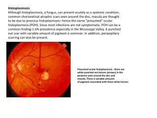
HISTOPLASMOSIS (Histoplasma capsulatum) Histoplasmosis is a systemic disease, mostly of the reticulo-endothelial system, manifesting itself in the bone marrow, lungs, liver, and the spleen. In fact, hepatosplenomegaly is the primary sign in children, while in adults, histoplasmosis more commonly appears as pulmonary disease. This is one of the most common fungal infections. The ecological niche of H. capsulatum is in blackbird roosts, chicken houses and bat guano. Typically, a patient will have spread chicken manure around his garden and 3 weeks later will develop pulmonary infection. Histoplasmosis is a significant occupational disease in bat caves in Mexico when workers harvest the guano for fertilizer. In the endemic area the majority of patients who develop histoplasmosis (95%) are asymptomatic. The diagnosis is made from their history, serologic testing or skin test.
Systemic Mycoses Five fungi are included in this group: Histoplasma capsulatum Blastomyces dermatitidis Paracoccidioides barasiliensis Coccidioides immitis Cryptococcus neoformans Four of these pathogens [H. capsulatum, B dermatitidis, P. barasiliensis and C. immitis] are dimorphic. They grow as filamentous molds as saprobes and in culture at 25 ゚C; when they infect humans or are cultured at 37 ゚C , they transform to a unicellular morphology.
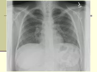
In the patients who are clinically ill, histoplasmosis generally occurs in one of three forms: acute pulmonary, chronic pulmonary or disseminated. There is generally complete recovery from the acute pulmonary form (another "flu-like" illness). However, if untreated, the disseminated form of disease is usually fatal. Patients will first notice shortness of breath and a cough which becomes productive. The sputum may be purulent or bloody. Patients will become anorexic and lose weight. They have night sweats. This again sounds like tuberculosis, and the lung X- ray also looks like tuberculosis, but today radiologists can distinguish between these diseases on the chest film (histoplasmosis usually appears as bilateral interstitial infiltrates).The skin test is NOT used for diagnostic purposes, because itinterferes with serological tests. Skin tests are used for epidemiological surveys.
Clinical specimens sent to the lab depend on the presentation of the disease: Sputum or Bronchial alveolar lavage, if it is pulmonary disease, or Biopsy material from the diseased organ. Bone marrow is an excellent source of the fungus, which tends to grow in the reticulo-endothelial system. Peripheral blood is also a source of visualizing the organism histologically. The yeast is usually found in monocytes or in PMN's. Many times an astute medical technologist performing a white blood cell count will be the first one to make the diagnosis of histoplasmosis. In peripheral blood, H. capsulatum appears as a small yeast about 5-6 µ in diameter. (Blastomyces is 12 to 15 µ). Gastric washings are also a source of H. capsulatum as people with pulmonary disease produce sputum and frequently swallow their sputum.
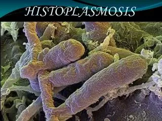
Mycology When it is grown on Sabouraud dextrose agar at 25ºC, it appears as a white, cottony mycelium after 2 to 3 weeks. The mold phase of H. capsulatum is characterized by thin, branching, septate hyphae that produce microconidia and a very distinct spore called a tuberculate macroconidium. The tubercles are diagnostic, however there are some non-pathogens which appear similar. Grown at 37ºC the budding yeast form appears. It is a white to tan colony. The yeast cell is 5-6 µ in diameter and slightly oval in shape and found exclusively within macrophages. To confirm the diagnosis, one must convert the organism from yeast to mycelium or vice-versa or use the DNA probe.
Serological Test • Serology for histoplasmosis is a little more complicated than for other mycoses, but it • provides more information than blastomycosis serology. • There are 4 tests: • Latex agglutination • Complement Fixation • Immunodiffusion • EIA
Each of these serological tests has different characteristics that make them useful. The latex agglutination test is a very simple test involving agglutination in a test tube. The Ab is fairly specific and rises early in the disease (in the first 2 weeks), and disappears in about 3 months. Thecomplement fixation ( C-F) test is like the one for blastomycosis,except there are 2 antigens, one to the yeast form of the organism and the other to the mycelial form. Some patients react to one form and not the other, while some individuals react to both. The reason for the different responses is not clear. Onedisadvantageis thatcomplement fixing antibody develops late in the disease, about 2 to 3 months after onset. A seconddisadvantage is that itcross reacts with other mycotic infections.
An advantage of the C-F test is that it is quantitative, so the physician can follow the course of the disease by observing the titer of several samples. The interpretation of the immunodiffusion test is a little more complicated than with blastomycosis because there are two bands which may appear. An H band indicates active disease and will appear in 2 to 3 weeks. An M band can indicate past or present disease, or result from a skin test. This is one reason why skin tests are not used for diagnosis because they can interfere with other tests. Skin tests will also affect the complement fixation test.
Recently, a radioimmunoassay for histoplasma polysaccharide antigen has been developed. This is a proprietary test so the evaluation of the results have been questioned. Treatment The drug of choice (DOC) is amphotericin B, with all its side effects. Itraconazole and Voriconazole is now also being used.