Download

1 / 95
970 likes | 1.27k Vues
David Clunie ( dclunie@dclunie.com ) PixelMed Publishing. DICOM WG 30 Small Animal Imaging DICOM Training Introduction and Basic Concepts. DICOM – Learning Objectives. Brief history File formats and Storage S ervices Data S et encoding principles Transfer Syntaxes

E N D
David Clunie (dclunie@dclunie.com) PixelMed Publishing DICOM WG 30 Small Animal ImagingDICOM TrainingIntroduction and Basic Concepts
DICOM – Learning Objectives • Brief history • File formats and Storage Services • Data Set encoding principles • Transfer Syntaxes • Information Object Definitions • Storage SOP Classes • Other DICOM services than storage • Conformance statements • Association Negotiation • DICOM Message Service Elements (DIMSE) • DICOM Upper Layer Protocol (DUL)
Who Needs to Know What? • User/customer • services and objects relevant to domain (esp. storage) • conformance statement interpretation and matching • Installer/integrator • + network addressing, more detail about services • +/- integration with non-imaging (IS, workflow) systems • Application designer/developer • detailed knowledge of (relevant) services, objects, attributes • uses toolkit to abstract (most) encoding/network details • Problem solver/troubleshooter • + data sets: interpret dumps and validation tool output • + network: DIMSE/DUL to interpret network logs/traces/dumps • Toolkit designer/developer • everything: objects, encoding, services, network • Standard writer: new modality • IOD structure, reusable modules, encoding limits, precedents
DICOM – Brief History • 1982 – 1stPACS Conference – session on standards • 1982 – AAPM Report 10 – Standard Format for Image Interchange • 1983 – ad hoc meeting between FDA, ACR & NEMA • 1983 – 1st meeting of ACR-NEMA “Digital Imaging and Communications Standards” Committee • 1985 – ACR-NEMA 300-1985 (“version 1.0”) issued • 1988 – ACR-NEMA 300-1988 (“version 2.0”) issued • 1990 – Inter-vendor testing of version 2.0 at Georgetown • 1992 – Trial of DICOM (“version 3.0) at RSNA • 1993 – DICOM 3.0 issued
DICOM – Brief History • 1982 – 1stPACS Conference – session on standards • 1982 – AAPM Report 10 – Standard Format for Image Interchange • 1983 – ad hoc meeting between FDA, ACR & NEMA • 1983 – 1st meeting of ACR-NEMA “Digital Imaging and Communications Standards” Committee • 1985 – ACR-NEMA 300-1985 (“version 1.0”) issued • 1985 – IEEE 802.3 Ethernet (based on 1976 Metcalfe) • 1986 – Aldus TIFF (version 3; prior versions drafts only) • 1987 – CompuServe GIF • 1988 – ACR-NEMA 300-1988 (“version 2.0”) issued • 1990 – Inter-vendor testing of version 2.0 at Georgetown • 1992 – Trial of DICOM (“version 3.0) at RSNA • 1992 – JPEG (ITU T.81; ISO 10918-1 1994) • 1993 – DICOM 3.0 issued
DICOM – Brief History • ACR-NEMA versions 1 and 2 • 50-pin 16 bit parallel interface • no network (assumed “network interface unit”) • layered • messages with commands and data • tag-value pairs • described patients, studies, images • described modality, acquisition, 3D position, etc.
DICOM – Brief History • ACR-NEMA versions 1 and 2 • 50-pin 16 bit parallel interface • no network (assumed “network interface unit”) • layered • messages with commands and data • tag-value pairs • described patients, studies, images • described modality, acquisition, 3D position, etc. • DICOM “3.0” • TCP/IP network protocol (and OSI semantics) • “object-oriented” description & conformance
DICOM – Scope • Implicit in the standard’s name: “Digital Image and Communications in Medicine” • Images and image-related information • All acquisition modalities • 1993: CR, CT, MR, US, NM, SC (DF and DV) • 2014: XA, XRF, DX, MG (+DBT), IO, PT, VL (Photo, Endo, Slide), WSI, OCT (OP,IV) • RT, SR (+CAD), waveforms, OP measurements and maps, surface scans, implants, segmentations • Storage + other services
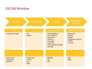
DICOM – Scope • Interchange of medical images • radiology, cardiology, pathology .... any ‘ology • network (TCP/IP) or media (CD, DVD, MOD) • Printing • grayscaleand color • network rather than point-to-point (TCP/IP) • Information System Integration • reports, worklists, performed procedures
Why does DICOM exist ? • Previous solutions closed (proprietary) • Real world requires open standards • heterogeneous modalities • heterogeneous workstations • heterogeneous archives, PACS, etc. • Consortium of • industry (NEMA) • users (ACR, ACC, CAP ...)
Why interchange images ? • Archiving • Interpretation • softcopy reading • filmless operation • Advanced processing • 3D visualization, quantitative analysis • fusion of images from multiple modalities • Teaching, research ...
Why (open) standards at all ? • Too many permutations of ... • Acquisition device vendors • general • GE, Siemens, Philips, Toshiba,Shimadzu, Hitachi, ... • special • Bruker, iTheraMedical, Mediso, PerkinElmer ... • Processing and archive device vendors • general • GE, Siemens, Philips, ... (+ open source) • special • InViCRO, ...
Why not existing standards ? • Didn’t “exist” when ACR-NEMA formed • Pure imaging standards (TIFF, etc.) • limited support for medical image types • don’t encode domain specific information • Other domains inappropriate • military, remote sensing, astronomical, etc. • ISO standards (e.g., IPI) never adopted • Medical standards that didn’t do images • HL7, MIB (IEEE 1073), etc. • Needed more services than just storage • query/retrieve, printing, workflow, etc.
DICOM – Why? • Domain-specific fields • patient identification & characteristics • device identification & characteristics • acquisition protocol parameters • study management identifiers, dates and times • Need to handle binary bulk pixel data • e.g., HL7 text based and no image support • Need to be extensible • evolving technology, private extensions • variable rather than “fixed” length headers
Key goals of DICOM • Support interoperability • NOT (necessarily) interfunctionality • WITHOUT defining (restricting) architecture • Define conformance • specific services and objects • documentation (Conformance Statement) • negotiation • Consensus standard • Voluntary compliance • Open (license fee free)
DICOM does NOT define: • PACS or IM Architecture • Picture Archiving Communications System • Image Management • Distributed Object Management • Radiology/Hospital Information System • Complete Electronic Medical Record Integrating the Health Care Enterprise (IHE) does define some aspects of architecture on top of DICOM, HL7, etc.
What is Interoperability? • Analogy of web server/browser: • Interconnectivity - both talk TCP/IP • Interoperability - both talk HTTP and HTML • Interfunctionality– approached, not guaranteed: • “versions” of HTML poorly controlled • layout and other behavior not constrained by HTML • optional/additional “features” • use of extensions (plug-ins, applets, scripts, stylesheets) • Not (always) sufficient for clinical needs • interoperability is necessary but not sufficient • reality check on user/customer expectations
DICOM and Interoperability • For example, conformance to DICOM • will guarantee network connection • will guarantee storage of MR image: • from Modality to Workstation • will NOT guarantee (but will facilitate): • Workstation will display image “correctly” • Workstation can perform analysis (e.g., diffusion tractography) • facilitated by mandatory attributes for: • identification, annotation, positioning, etc. IHE profiles define functionality,e.g., DIFF, PERF, MAMMO, Cardiac NM
DICOM and Interoperability • “Object-oriented” definition • data structures, e.g., MR image object • composite model of real world entities • patient, study, series • general image, specialized to MR image • services, e.g., image storage • together -> service/object pairs (SOP) • Roles (user or provider) (SCU or SCP) • Role + SOP Class -> Conformance
DICOM SOP Classes/Roles • MR scanner may say: • “I am an MR Image Storage Service Class User (SCU)” • Workstation may say: • “I am an MR Image Storage Service Class Provider (SCP) (amongst other things)”MR images may be transferred
DICOM SOP Classes/Roles • Angiography device may say: • “I am an XA Image Storage Service Class User (SCU)” • Workstation may say: • “I am not an XA Image Storage Service Class Provider (SCP) (though I do support other kinds of images like CT and MR)”This pair cannot transfer XA images
Why is DICOM so specific ? • For example, • MR Image • single frame, 12-16 bit grayscale image • MR acquisition - pulse sequence parameters • 3D patient relative co-ordinate/vector position • X-Ray Angiography Image • multi-frame, 8-10 bit grayscale image • XA acquisition - radiation/collimation/motion • dynamic C-arm/table relative positioning
DICOM SOP Classes/Roles • Workstation may say: • “I am a Basic Grayscale Print Management Meta SOP Class SCU” • Printer may say: • “I am a Basic Grayscale Print Management Meta SOP Class SCP”Images may be printed
DICOM SOP Classes/Roles • Ultrasound scanner may say: • “I am a Basic Color Print Management Meta SOP Class SCU” • Printer may say: • “I am only a Basic Grayscale Print Management Meta SOP Class SCP”This pair cannot print images
Modality is Important • Different acquisition characteristics • X-Ray-based v. not • cross-sectional v. projectional (planar) • transmissive v. emissive • structural v. functional • different mechanisms of contrast (endogenous or exogenous) • hybrids
Modality is Important • Different image characteristics • single or multiple channels (grayscale, color) • use of pseudo-color (palette color) • broader dynamic range than displayed • values may be > 8 bits in depth and signed • non-linear transformation (or linear window) • physical significance of pixel values (counts, HU, velocity) • physical significance of size (measurements)
Modality-specific DICOM IODs • Information Object Definition (IOD) • one (or more) for each modality • more if conformance reason for variants • “Composite” • multiple real-world entities in same IOD • “Object-oriented” (sort of) • “inherit” same (composite) real-world/information “model” • patient/study/procedure step/series/image/frame • re-use common general Modules • e.g., Patient, General Study, General Image Module • re-use common technology Modules • e.g., Contrast/Bolus, Frame of Reference, X-Ray Grid Module • “specialize” with modality-specific Modules • e.g., CT Image, MR Image Module
Image Plane Module Type: 1 – required, 2 – may be empty if unknown, 3 – optional
Encoding – Data Elements • Flat list of sorted unique “Data Elements” • IOD/Module structure is NOT encoded • Module Attributes encoded as Data Elements • Each Data Element is a Tag-Value pair • Tag-Type-Value triple with explicit type (VR) encoding • Data Element Tag: pair of 16 bit numbers • 16 bit “group number” (historical distinction) • 16 bit “element number” • encoded in binary, described in hexadecimal • e.g., (0008,0020) means “Study Date” • “meaning” is not encoded – need “data dictionary” to look them up (PS3.6) – i.e., not “self-describing”
Encoding – Data Elements • Value • Value Representation (VR) (“type”) • Value Length (16 or 32 bits; limits max size) • Value (even length padded; 16 bit parallel legacy) • VRs (“types”) • binary: US, SS, UL, SL, FL, FD, AT • bulk binary: OB, OW, OF, OD, UN (“unknown”) • numeric string: AS, DS, IS, DA, DT, TM • string: AE, CS, LO, PN, SH, UI • text: LT, ST,UT
Example MR Image Dataset (0x0008,0x0005) CS Specific Character Set VR=<CS> VL=<0x000a> <ISO_IR 100> (0x0008,0x0008) CS Image Type VR=<CS> VL=<0x0010> <ORIGINAL\PRIMARY> (0x0008,0x0016) UI SOP Class UID VR=<UI> VL=<0x001a> <1.2.840.10008.5.1.4.1.1.4> (0x0008,0x0018) UI SOP Instance UID VR=<UI> VL=<0x002e> <1.2.840.113619.2.1.2.1909421756.1.1.602501582> (0x0008,0x0020) DA Study Date VR=<DA> VL=<0x0008> <19890203> (0x0008,0x0021) DA Series Date VR=<DA> VL=<0x0008> <19890203> (0x0008,0x0022) DA Acquisition Date VR=<DA> VL=<0x0008> <19890203> (0x0008,0x0023) DA Image Date VR=<DA> VL=<0x0008> <19890203> (0x0008,0x0030) TM Study Time VR=<TM> VL=<0x0006> <092618> (0x0008,0x0031) TM Series Time VR=<TM> VL=<0x0006> <093221> (0x0008,0x0032) TM Acquisition Time VR=<TM> VL=<0x0006> <093302> (0x0008,0x0033) TM Image Time VR=<TM> VL=<0x0006> <093302> (0x0008,0x0050) SH Accession Number VR=<SH> VL=<0x0000> [] (0x0008,0x0060) CS Modality VR=<CS> VL=<0x0002> <MR> (0x0008,0x0070) LO Manufacturer VR=<LO> VL=<0x0012> <GE MEDICAL SYSTEMS> (0x0008,0x0080) LO Institution Name VR=<LO> VL=<0x001c> <THOMAS JEFF UNIVHOSPITAL MRI> (0x0008,0x0090) PN Referring Physician's Name VR=<PN> VL=<0x0004> <HUME> (0x0008,0x1010) SH Station Name VR=<SH> VL=<0x0008> <FOR.IC0 > (0x0008,0x1030) LO Study Description VR=<LO> VL=<0x0004> <KNEE> (0x0008,0x103e) LO Series Description VR=<LO> VL=<0x0006> <COR T2> (0x0008,0x1060) PN Name of Physician(s) Reading Study VR=<PN> VL=<0x0004> <BODY> (0x0008,0x1070) PN Operator's Name VR=<PN> VL=<0x0002> <RB> (0x0008,0x1090) LO Manufacturer's Model Name VR=<LO> VL=<0x000e> <GENESIS_SIGNA > (0x0010,0x0010) PN Patient's Name VR=<PN> VL=<0x000c> <* GRX KNEE *> (0x0010,0x0020) LO Patient's ID VR=<LO> VL=<0x0006> <RSNA2 > (0x0010,0x0030) DA Patient's Birth Date VR=<DA> VL=<0x0000> [] (0x0010,0x0040) CS Patient's Sex VR=<CS> VL=<0x0002> <M > (0x0010,0x1010) AS Patient's Age VR=<AS> VL=<0x0004> <034Y> (0x0010,0x1030) DS Patient's Weight VR=<DS> VL=<0x000a> < 90.718000> (0x0010,0x21b0) LT Additional Patient History VR=<LT> VL=<0x0008> <R/O TEAR> (0x0018,0x0020) CS Scanning Sequence VR=<CS> VL=<0x0002> <SE> (0x0018,0x0021) CS Sequence Variant VR=<CS> VL=<0x0004> <OSP > (0x0018,0x0022) CS Scan Options VR=<CS> VL=<0x0004> <NPW > (0x0018,0x0023) CS MR Acquisition Type VR=<CS> VL=<0x0002> <2D> (0x0018,0x0025) CS Angio Flag VR=<CS> VL=<0x0002> <N > (0x0018,0x0050) DS Slice Thickness VR=<DS> VL=<0x0008> <5.000000> (0x0018,0x0080) DS Repetition Time VR=<DS> VL=<0x000c> < 2000.000000> (0x0018,0x0081) DS Echo Time VR=<DS> VL=<0x000a> < 20.000000> (0x0018,0x0082) DS Inversion Time VR=<DS> VL=<0x0008> <0.000000> (0x0018,0x0083) DS Number of Averages VR=<DS> VL=<0x0008> <0.500000> (0x0018,0x0084) DS Imaging Frequency VR=<DS> VL=<0x0010> < 638746840.00000> (0x0018,0x0085) SH Imaged Nucleus VR=<SH> VL=<0x0002> <H1> (0x0018,0x0086) IS Echo Number(s) VR=<IS> VL=<0x0002> < 1> (0x0018,0x0087) DS Magnetic Field Strength VR=<DS> VL=<0x0006> <15000 > (0x0018,0x0088) DS Spacing Between Slices VR=<DS> VL=<0x0008> <6.000000> (0x0018,0x0091) IS Echo Train Length VR=<IS> VL=<0x0002> <0 > (0x0018,0x0093) DS Percent Sampling VR=<DS> VL=<0x000a> < 53.125000> (0x0018,0x0094) DS Percent Phase Field of View VR=<DS> VL=<0x000a> <100.000000> (0x0018,0x1088) IS Heart Rate VR=<IS> VL=<0x0002> <0 > (0x0018,0x1090) IS Cardiac Number of Images VR=<IS> VL=<0x0002> <0 > (0x0018,0x1094) IS Trigger Window VR=<IS> VL=<0x0002> <10> (0x0018,0x1100) DS Reconstruction Diameter VR=<DS> VL=<0x000a> <140.000000> (0x0018,0x1314) DS Flip Angle VR=<DS> VL=<0x0002> <0 > (0x0018,0x1315) CS Variable Flip Angle Flag VR=<CS> VL=<0x0002> <N > (0x0018,0x1316) DS SAR VR=<DS> VL=<0x0008> <0.052993> (0x0018,0x5100) CS Patient Position VR=<CS> VL=<0x0004> <FFS > (0x0020,0x000d) UI Study Instance UID VR=<UI> VL=<0x0028> <1.2.840.113619.2.1.2.139348932.602501178> (0x0020,0x000e) UI Series Instance UID VR=<UI> VL=<0x002a> <1.2.840.113619.2.1.2.596272627.1.602501541> (0x0020,0x0010) SH Study ID VR=<SH> VL=<0x0002> <2 > (0x0020,0x0011) IS Series Number VR=<IS> VL=<0x0002> < 1> (0x0020,0x0012) IS Acquisition Number VR=<IS> VL=<0x0002> < 0> (0x0020,0x0013) IS Image Number VR=<IS> VL=<0x0002> < 1> (0x0020,0x0032) DS Image Position (Patient) VR=<DS> VL=<0x0020> <-70.000000\ 18.000000\ 75.000000> (0x0020,0x0037) DS Image Orientation (Patient) VR=<DS> VL=<0x0038> < 1.000000\0.000000\0.000000\0.000000\0.000000\ -1.000000> (0x0020,0x0052) UI Frame of Reference UID VR=<UI> VL=<0x002c> <1.2.840.113619.2.1.2.596272627.1.602501541.0> (0x0020,0x0060) CS Laterality VR=<CS> VL=<0x0000> [] (0x0020,0x0110) DS Temporal Resolution VR=<DS> VL=<0x000a> <1120403456> (0x0020,0x1040) LO Position Reference Indicator VR=<LO> VL=<0x0002> <KN> (0x0020,0x1041) DS Slice Location VR=<DS> VL=<0x000e> <-18.0000000000> (0x0028,0x0002) US Samples per Pixel VR=<US> VL=<0x0002> [0x01] (0x0028,0x0004) CS Photometric Interpretation VR=<CS> VL=<0x000c> <MONOCHROME2 > (0x0028,0x0010) US Rows VR=<US> VL=<0x0002> [0x100] (0x0028,0x0011) US Columns VR=<US> VL=<0x0002> [0x100] (0x0028,0x0030) DS Pixel Spacing VR=<DS> VL=<0x0012> < 0.546875\0.546875> (0x0028,0x0100) US Bits Allocated VR=<US> VL=<0x0002> [0x10] (0x0028,0x0101) US Bits Stored VR=<US> VL=<0x0002> [0x10] (0x0028,0x0102) US High Bit VR=<US> VL=<0x0002> [0x0f] (0x0028,0x0103) US Pixel Representation VR=<US> VL=<0x0002> [0x01] (0x0028,0x0120) XS Pixel Padding Value VR=<SS> VL=<0x0002> [0x00] (0x7fe0,0x0010) OX Pixel Data VR=<OW> VL=<0x20000> []
Encoding – Sequences • Sequence (SQ) VR allows “nesting” of lists • Data Element with an SQ VR • no “value” field per se • may have a fixed or undefined length • zero or more Sequence Items • Sequence Delimiter tag (if was undefined length) • Sequence Item • Item tag (may have a fixed or undefined length) • list of (sorted, unique) data elements • Item Delimiter tag (if was undefined length)
Dump Tool Output for Sequence %item (0x0040,0xa010) Relationship Type <HAS OBS CONTEXT> (0x0040,0xa040) Value Type <PNAME > (0x0040,0xa043) Concept Name Code Sequence %item (0x0008,0x0100) Code Value <000555> (0x0008,0x0102) Coding Scheme Designator <LNdemo> (0x0008,0x0104) Code Meaning <Recording 0bserver> %enditem %endseq (0x0040,0xa123) Person Name <Smith^John^^Dr^ > %enditem
Actual Binary Encoding ... (0x0028,0x0002) Samples per Pixel VR=<US> VL=<0x0002> [0x0001] (0x0028,0x0004) Photometric Interpretation VR=<CS> VL=<0x000c> <MONOCHROME2 > (0x0028,0x0008) Number of Frames VR=<IS> VL=<0x0004> <124 > (0x0028,0x0010) Rows VR=<US> VL=<0x0002> [0x0100] (0x0028,0x0011) Columns VR=<US> VL=<0x0002> [0x0100] (0x0028,0x0100) Bits Allocated VR=<US> VL=<0x0002> [0x0010] (0x0028,0x0101) Bits Stored VR=<US> VL=<0x0002> [0x0010] (0x0028,0x0102) High Bit VR=<US> VL=<0x0002> [0x000f] ... ... 00000560 .. .. .. .. .. .. 28 00 02 00 55 53 02 00 01 00 |..........US....| 00000570 28 00 04 00 43 53 0c 00 4d 4f 4e 4f 43 48 52 4f |(...CS..MONOCHRO| 00000580 4d 45 32 20 28 00 08 00 49 53 04 00 31 32 34 20 |ME2 (...IS..124 | 00000590 28 00 10 00 55 53 02 00 00 01 28 00 11 00 55 53 |(...US....(...US| 000005a0 02 00 00 01 28 00 00 01 55 53 02 00 10 00 28 00 |....(...US....(.| 000005b0 01 01 55 53 02 00 10 00 28 00 02 01 55 53 02 00 |..US....(...US..| ... *names shown in test tool dump are not actually encoded
You Need a Libraryor Toolkit • You can hand-code reading & writing • But: • legal variations in encoding (Transfer Syntax) • sequence handling is non-trivial • need a “data dictionary” to understand tags • need an “IOD” library to map flat list of data elements as encoded to/from IODs/Modules (or “classes”) • need utilities for dumping, testing, editing, etc. • Not to mention files, services, protocols! • Fortunately, are available for all platforms
Private Data Elements • Odd group numbers are all private • (gggg,00xx) is a private creator string • (gggg,xxyy) is the block defined by that creator (0019,0010) “David’s Stuff” (0019,0011) “Harry’s Stuff” (0019,1001) 1st of david’sprivate data elements (0019,1002) 2nd of david’sprivate data elements … (0019,1101) 1st of harry’sprivate data elements (0019,1102) 2nd of harry’sprivate data elements …
DICOM – Is it a “File Format”? • Yes and no • since 1995 – a formal “file format” has been defined (PS3.10 aka. “part 10”) • but, it applies only to files on specified media (e.g., CD) with DICOMDIR (directory) (“File Set”) • strictly speaking, no standard conformance requirements for files “by themselves” • In practice • esp. for research and testing, “DICOM files” are often stored and exchanged informally • even old ACR-NEMA data sets were stored in files and interchanged
File Format v. Data Set • Data Set • “an instance of a real world Information Object” • “constructed of Data Elements” • is what is exchanged on network using the C-STORE “command” • File Format • “a means to encapsulate in a file the Data Set” • “byte stream of the Data Set is placed into the file after the DICOM File Meta Information” • “each file contains a single SOP Instance”
Data Set Data Set Data Elements
Data Set Data Set Data Elements Pixel Data Element