Download
1 / 24
530 likes | 1.51k Vues
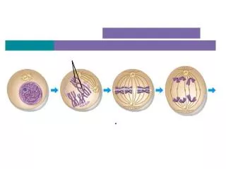
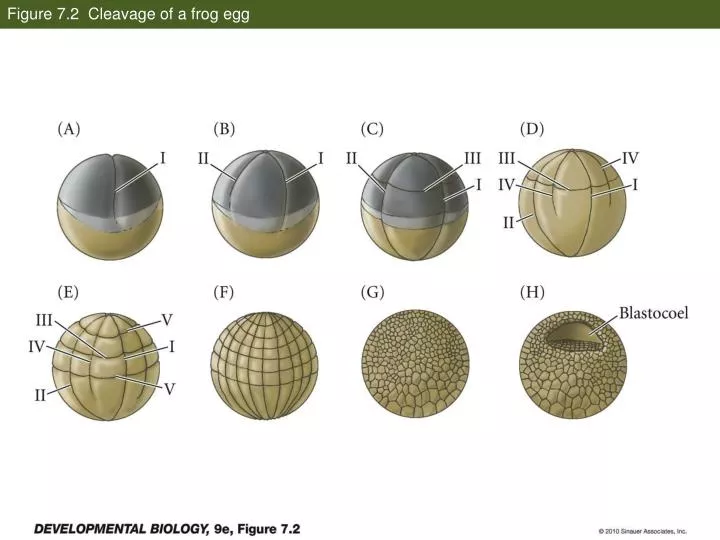
Figure 7.2 Cleavage of a frog egg. Figure 7.3 Scanning electron micrographs of frog egg cleavage. Figure 7.5 Fate maps of the Xenopus laevis blastula exterior (A) and interior (B) . Figure 7.6 Cell movements during frog gastrulation (Part 1).

E N D
Figure 7.3 Scanning electron micrographs of frog egg cleavage
Figure 7.5 Fate maps of the Xenopus laevis blastula exterior (A) and interior (B)
Figure 7.7 Surface view of an early dorsal blastopore lip of Xenopus
Figure 7.11 Protocadherin expression separates axial and paraxial mesoderm
Figure 12.1 Mesodermal development in frog and chick embryos
Figure 8.15 Development of a human embryo from fertilization to implantation
Figure 8.23 Amnion structure and cell movements during human gastrulation
Figure 12.1 Mesodermal development in frog and chick embryos
Primitive streak stage (7.5 dpc) T/Bra tbx6 Chapman DL, Agulnik I, Hancock S, Silver LM, Papaioannou VE. Tbx6, a mouse T-Box gene implicated in paraxial mesoderm formation at gastrulation. DevBiol. 1996 Dec 15;180(2):534-42.
Summary Chapman DL, Papaioannou VE. Three neural tubes in mouse embryos with mutations in the T-box gene Tbx6.Nature. 1998 Feb 12;391(6668):695-7.