Download
1 / 6
60 likes | 243 Vues
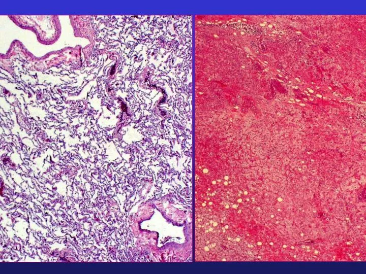
Final Diagnosis: Lung Mass, Right (wedge excision): - Metastatic myxoid Liposarcoma, grade 2 of 4. Note: This tumor has an appearance similar to the previously sampled material (SXX-XXXXX). The tumor does not appear to have increased in grade.

E N D
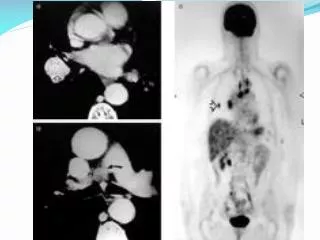
Final Diagnosis: Lung Mass, Right (wedge excision): - Metastatic myxoid Liposarcoma, grade 2 of 4. Note: This tumor has an appearance similar to the previously sampled material (SXX-XXXXX). The tumor does not appear to have increased in grade.
Metastatic patterns of extremity myxoid liposarcoma and their outcome.Estourgie SH, Nielsen GP, Ott MJ.J Surg Oncol 2002 Jun;80(2):89-93 • School of Medicine, University of Utrecht, Utrecht, The Netherlands. • BACKGROUND AND OBJECTIVES: Extremity myxoid liposarcomas have a unique extrapulmonary metastatic potential. We studied the metastatic pattern of extremity liposarcomas to determine what types of posttreatment imaging may be of value in the follow-up these patients. METHODS: Twenty-two patients from a total of 128 patients with primary extremity liposarcoma were treated at a tertiary care institution for subsequent metastases from January 1981 to January 2000. Median follow-up was 45 months (range: 6-270 months). Data on these patients was prospectively collected and then retrospectively analyzed for effect of metastatic pattern and treatment on outcome. RESULTS: Of these 22 patients, extrapulmonary metastases developed in 10, combined pulmonary and extrapulmonary metastases developed in 6, and isolated pulmonary metastases developed in 6.Of the 16 patients with extrapulmonary metastases, 13 were of the myxoid subtype. Of the 49 patients with extremity myxoid liposarcomas, metastases developed in 14 (29%). The most common sites of metastases among these 14 patients include: the retroperitoneum, 10 patients (71)%; intra-abdominal extra-hepatic, 7 patients (50%); spinal/paraspinal, 6 patients (43%). Only 3 of the patients are alive and disease free and all 3 of these patients are from the subgroup of 10 patients with only extra-pulmonary metastases (2 intra-abdominal and 1 retroperitoneal). CONCLUSIONS: Extremity myxoid liposarcomas have an unusually high predilection for extra-pulmonary metastases, frequently without any pulmonary metastases. After treatment of the primary tumor, these patients should be followed with periodic chest X-ray and abdominal/pelvic computed tomography (CT) scans. Any back or neurologic complaints should prompt additional imaging of the appropriate spinal area. Consideration should be given to surgical and adjuvant treatment of metastatic disease when appropriate.