Download
1 / 53
530 likes | 887 Vues
Ovarian hormones can influence motor activity. review main components of the motor system consider role of hormones in altering motor responses by acting on neurons within the basal ganglia and cerebellum

E N D
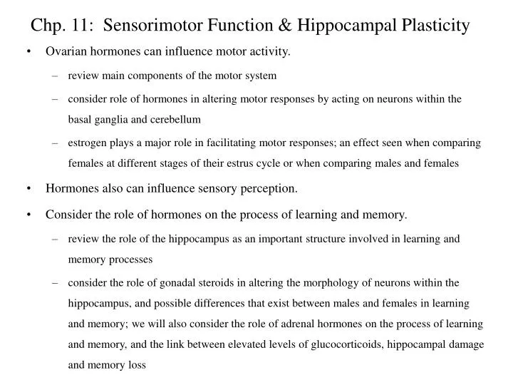
Ovarian hormones can influence motor activity. review main components of the motor system consider role of hormones in altering motor responses by acting on neurons within the basal ganglia and cerebellum estrogen plays a major role in facilitating motor responses; an effect seen when comparing females at different stages of their estrus cycle or when comparing males and females Hormones also can influence sensory perception. Consider the role of hormones on the process of learning and memory. review the role of the hippocampus as an important structure involved in learning and memory processes consider the role of gonadal steroids in altering the morphology of neurons within the hippocampus, and possible differences that exist between males and females in learning and memory; we will also consider the role of adrenal hormones on the process of learning and memory, and the link between elevated levels of glucocorticoids, hippocampal damage and memory loss Chp. 11: Sensorimotor Function & Hippocampal Plasticity
The motor system can be divided into two groups of circuits: pyramidal system: consists of pyramidal neurons within the cerebral cortex that project to lower motorneurons in the brainstem and spinal cord to control voluntary movement several cortical areas are involved in the initiation of movement: neuronal activity within supplementary motor area and premotor cortex precedes activity within the primary motor cortex neuronal activity in the primary motor cortex is associated with the initiation of movement pyramidal neurons within the primary motor cortex project to, and activate, lower motorneurons in the brainstem and spinal cord lower motor neurons innervate skeletal muscle controlling contraction of various muscle groups and the movement of body parts (e.g., movement of arm or chewing) Motor System Supplementary Motor Area Premotor Cortex Primary Motor Cortex initiation of movement
The motor system can be divided into two groups of circuits: extrapyramidal system: composed of all other projection pathways that influence motor control: basal ganglia cerebellum groups of neurons within the brainstem that send projections into the spinal cord neurons within the basal ganglia and cerebellum are interconnected with the cerebral cortex through a series of feedback loops--one way in which the basal ganglia and cerebellum can influence motor responses in addition, components of the basal ganglia have been linked to cognitive processes (memory) Motor System
Basal Ganglia: the basal ganglia include: caudate nucleus, putamen and globus pallidus in humans, the caudate nucleus and putamen are typically segregated; in lower mammals (like the rat), the caudate nucleus and putamen are combined into one structure striatum is a term used to refer to both the caudate nucleus and putamen corpus striatum refers to the caudate nucleus, putamen and globus pallidus two additional brain regions are interconnected with the basal ganglia--subthalamic nucleus and substantia nigra the basal ganglia forms a variety of interconnected loops with the subthalamic nucleus, substantia nigra, thalamus and cerebral cortex bottom line: basal ganglia (and associated brain regions) receives input from sensory and motor cortices, it processes and integrates the information, and then sends the output to supplementary and premotor cortices to control motor activity Motor System
The brain is organized bilaterally--with similar brain structures present on right and left sides. L R Primary Motor Cortex Striatum Substantia nigra provides an important source of dopamine to the striatum Substantia nigra In most instances, motor control is contralateral such that the right primary motor cortex controls movements on the left side of the body. control of movement on the left side of the body
In humans, two clinical syndromes have provided insight into the basic function of the basal ganglia: Parkinson’s disease and Huntington’s disease Parkinson’s disease: dopamine neurons within the substantia nigra degenerate leading to increased inhibition of motor activity individuals with this disorder show several symptoms including: bradykinesia--a reduction in the speed of movements difficulty initiating and stopping movements development of resting tremor--a regular involuntary, oscillatory movement of a body part, usually hands and extremities Motor System
In humans, two clinical syndromes have provided insight into the basic function of the basal ganglia: Parkinson’s disease and Huntington’s disease Huntington’s disease: degeneration of neurons within the striatum including neurons that synthesize the neurotransmitters GABA and acetylcholine effect of this cell loss is disinhibition of motor activity (increased activity) individuals with this disorder show several symptoms including: progressive dementia--cognitive deficits choreiform movements--rapid, irrregular flow of motion associated with fingers, arms and facial muscles; effects can include: “piano-playing” fexion-extension movements of the fingers, elevation and depression of the shoulders and hips, crossing and uncrossing of the legs, and grimacing movements of the face Huntington’s disease is a hereditary disease; onset of symptoms occurs during the third or fourth decade of life (30s and 40s) Motor System
In rodents, dopaminergic projections from the substantia nigra to the striatum have been implicated in motoric function: locomotion stereotyped behavior rotational behavior postural asymmetries These effects of dopamine are associated with dopaminergic receptors within the striatum; several subtypes are present: subtypes: D1 dopamine receptors and D2 dopamine receptors Estrogen plays an important role in facilitating dopamine neurotransmission within the striatum which leads to selective increases in motoric function: changes in motoric function in females at different stages of the estrus cycle sex differences in motor function Motor System
Different types of drugs have been used to study the effects of dopamine activity in the striatum: apomorphine--dopamine agonist that binds to dopamine receptors amphetamine--drug that stimulates release of dopamine from nerve terminals in the striatum; secondarily, then the released dopamine will bind to dopamine receptors haloperidol--dopamine receptor antagonist that acts by blocking dopamine receptors (blocks the ability of dopamine to bind to its receptor) The administration of apomorphine or amphetamine increases dopamine activity within the brain (including the striatum); two main motoric effects are produced: first, there is an increase in locomotion and exploratory behavior second, there is an increase in the display of stereotyped behaviors--repetitive movements of head, whiskers and forelimbs; these repetitive movements can include: chewing movements, excessive sniffing, up/down movements of the head, and so on Motor System
The administration of apomorphine or amphetamine can also induce rotational behavior; this phenomenon is usually studied in animals in which the nigrostriatal dopamine system has been damaged unilaterally. Depending on the drug that is given, animals will turn in a particular direction. Motor System Fewer dopamine terminals in the striatum on the lesioned side; less dopamine available for release. L R Striatum Selectively destroy dopamine neurons by administering a neurotoxin to the substantia nigra on one side (6-hydroxydopamine) Substantia nigra
Motor System Striatum Effect animal turns toward the lesion (away from striatum that has the greatest activity) increase in dopamine activity on intact side (reflects release) if you administer amphetamine animal turns away from the lesion (away from striatum that has the greatest activity) increase in dopamine activity on both sides (dopamine receptor activation) if you administer apomorphine receptor supersensitivity
Amphetamine stimulates dopamine release; more dopamine will be released on the intact side (greater activity); animal will turn toward lesion. Apomorphine stimulates dopamine receptors; reduced levels of dopamine on the lesioned side leads to an increase in dopamine receptors; more dopamine receptors will be activated on lesioned side (greater activity); animal will turn away from lesion. Motor System amphetamine Decreased levels of dopamine produce a compensatory increase in dopamine receptors in the striatum on the lesioned side. Turning apomorphine R L Striatum Dopamine receptor Substantia nigra
Bottom line: changes in gonadal steroids that occur during the estrus cycle (primarily the rise in estrogen) stimulates release of dopamine within the striatum that leads to alterations in the behavior of the female rat. estrogen levels are elevated during late proestrus-early estrus (prior to, and during the start of behavioral estrus and ovulation); estrogen levels are lower at other times (e.g., diestrus) dopamine synthesis and release within the striatum is greatest during estrus administration of amphetamine can stimulate greater release of dopamine in the striatum of female rats in estrus in comparison to females in diestrus; this can be seen in tissue slices of striatum that are placed into a tissue chamber and perfused with amphetamine; this can also be seen in freely moving rats using microdialysisto sample dopamine release within the striatum after administration of amphetamine administration of amphetamine produced greater levels of stereotyped behavior (such as sniffing and head and forelimb movements) in female rats in estrus in comparison to those in diestrus Motor System
administration of amphetamine also produced greater amounts of rotational behavior in females during estrus in comparison to females in diestrus removal of the ovaries (ovariectomy) reduces estrogen levels in female rats and will decrease stereotypy and rotational behavior induced by drugs associated with this decline in behavior is a decrease in the release of dopamine by amphetamine in ovariectomized females administration of estrogen to ovariectomized females will enhance dopamine-related behaviors and increase dopamine release within the striatum in response to amphetamine Motor System
The effects of estrogen on striatal activity can also be seen in spontaneous behaviors: you can train female rats to walk across a narrow beam suspended about 3 feet above the floor; task that reflects sensorimotor coordination you can analyze how well the female does on this task by examining the accuracy of foot placement on the beam--if the foot was placed on top of the narrow beam--”correct”, if the foot slipped off the top or grabbed onto the side--”footfault” Does performance on this task change over the estrus cycle? YES--the number of footfaults decrease during estrus; the female rat performs better on task when estrogen levels are high you can reproduce this effect by administering estrogen directly into the striatum Motor System implant cholesterol into striatum (control) no effect on # footfaults OVX female rats train females to walk on beam significant decline in # footfaults implant estrogen into striatum
How does estrogen induce its effects at the level of the striatum? its is presently unknown there are few neurons in the striatum that accumulate estrogen (few estrogen receptors) effects of estrogen may be nontraditional, that is, estrogen may act at the membrane of nerve terminals to enhance dopamine release versus control of gene transcription; there is evidence for rapid effects of gonadal steroids on membranes (but we don’t know, in most cases, how these rapid effects occur) it is possible that estrogen may alter other neurocircuits that project to the striatum and that regulate dopamine release (e.g., frontal cortex) it is also possible that estrogen may influence the amount of dopamine available for release; there are dopamine neurons within the substantia nigra that possess estrogen receptors however, these latter observations do not explain how estrogen implants within the striatum can alter dopamine release and behavior Motor System
In addition to differences in the behavior of female rats during different days of the estrus cycle, there are sex differences in the activation of motoric responses by drugs male rats show lower rates of rotational behavior, locomotor activity, and stereotypy in response to amphetamine than do female rats in estrus male rats also show lower level of dopamine release in response to amphetamine than do female rats in estrus castration of male rats has no effect on motoric responses nor on striatal dopamine release in contrast, ovariectomy in female rats significantly decreases amphetamine-induced dopamine release in the striatum and amphetamine-stimulated motoric behaviors important link between estrogen and dopamine-responsiveness in females Motor System
There are also differences between males and females in a more natural setting--open-field activity test. In the open-field activity test, a rat is placed in a large open testing arena and the amount of time spent walking around (ambulation) and rearing can be determined females ambulate and rear more than males ovariectomy decreases these responses, while castration has no effect in males The relationship between estrogen and increases motoric responses in the adult is associated with organizational and activation effects: androgens/estrogens need to be low during perinatal development for feminization of this behavioral response (organizational effect) increases in estrogen that occur in females during estrus cycle are needed to produce high levels of open-field activity in the adult The significance of estrogen-stimulated motoric responses may be associated with finding a mate and/or motoric responses involved with mating. Motor System
Cerebellum: the cerebellum receives sensory and motor input, processes the information, and then sends its output (via Purkinje cells) to deep cerebellar nuclei which integrate the input with other motor control systems bottom line: cerebellum is involved primarily in controlling the timing and pattern of muscles activated during movement, postural support and maintenance of muscle tone Purkinje cell firing is correlated with movement (electrophysiological studies) gonadal steroids have been shown to modulate the firing of Purkinje cells during movement: estrogen increases firing rate of Purkinje cells progesterone decreases the firing rate of Purkinje cells Motor System
Cerebellum: it has been suggested that the rise in estrogen followed by the rise in progesterone may play a role in sensorimotor gating in the cerebellum--influencing how the cerebellum responds to sensorimotor input and its role in controlling motor output the increases in estrogen and proesterone occur during late proestrus to early estrus and are associated with changes in proceptive and receptive (lordosis) responses; it is possible that changes occurring in Purkinje cell firing rates are associated with proceptive and receptive behaviors; although, how this occurs is not known of interest, progesterone can bind to GABA-A receptors to potentiate the effects of GABA at its receptor GABA is an inhibitory neurotransmitter that acts to increase chloride (Cl-) conductance into the cell, with a net effect of increased inhibition the binding of progesterone to the GABA-A receptor is thought to mediate the decrease in Purkinje cell firing that occurs when progesterone levels are high Motor System
In addition to hormonal effects on motor responses, hormones can also influence sensory perception. the vomeronasal organ mediates detection of pheromones that stimulate various responses such as activation of sex behavior or preferences for odors on gonadally-intact conspecifics gonadectomy decreases these responses administration of testosterone to males, and estrogen to females, can restore these responses There is also evidence that hormones can influence taste and pain sensitivity. female rats show a greater preference for sweet tastes and salt solutions than males stress-induced analgesia (opioid-dependent form) is greater in males than in females female rats are more responsive to electric footshock than males; during footshock, females respond to lower intensities of shock (lower pain thresholds) and react more quickly (shorter escape latencies) Sensory Perception
NEXT SECTION
Major cellular components: dentate gyrus (major source of inputs) Ammon’s horn: fields CA1, CA2, CA3/CA4 (integrative role) subiculum (major source of efferents) Major pathways and connections: hippocampus receives highly processed sensory information about internal and external events: perforant pathway: hippocampus is reciprocally connected to the entorhinal cortex; entorhinal cortex has connections with other corticial areas (visual, auditory information) fimbria-fornix: hippcampus is also reciprocally connected to septum, thalamus and hypothalamus several pathways that allow for intrahippocampal connections: commissural connections: neuronal connections made between neurons in two hippocampi Schaffer collaterals: connections between neurons made on one side of hippocampus Hippocampus
Hippocampus plays an important role in two main processes: learning and memory especially tasks that involve processes of spatial cues evidence for sex differences in hippocampal structure also evidence for sex differences in the performance of spatial tasks; gonadal steroids have been implicated in organizational and activational effects on performance in the adult, changes in hippocampal structure accompany hormone changes during estrus “brake” on HPA axis hippocampus possesses mineralocorticoid and glucocorticoid receptors mineralocorticoid receptors are linked to circadian changes in HPA axis glucocorticoid receptors are linked to terminating a stress response chronic exposure to glucocorticoids can damage the hippocampus leading to higher levels of glucocorticoids, more hippocampal damage, and so on; damage to the hippocampus has been linked to memory deficits Hippocampus
Patient H.M.: H.M. suffered from intractable epilepsy (epileptic seizures) an epileptic seizure means that a large collection of neurons in the brain discharge in abnormal synchrony--seizures can be focal that spread throughout cortex or generalized, and may involve loss of consciousness as well as contraction of groups of skeletal muscle intractable means that his epileptic seizures were resistant to treatment to stop his epileptic seizures, heunderwent bilateral hippocampectomy--bilateral removal of his hippocampi following surgery: GOOD NEWS: his epilepsy stopped BAD NEWS: while he could remember events early in his life, he could not remember events just prior to surgery (mild form of “retrograde amnesia”), and he was unable to form new memories (“anterograde amnesia:) Hippocampus
Patient H.M.: these events in Patient H.M. highlight the important role that the hippocampus plays in the processes of learning and memory mild form of retrograde amnesia and anterograde amnesia indicates that the hippocampus plays an important role in learning and in the formation of short-term memory (working memory) however, the ability of H.M. to remember early events in his life indicates that the hippocampus is not the location where long-term memories are stored Hippocampus
What processes have been implicated in learning and memory? long-term potentiation (LTP) an increase in neural activity at particular synapses will “strengthen” those synapses this strengthening process involves an in crease in synaptic efficacy which simply means that a greater synaptic response will be produced in response to a given input response is believed to last from hours to days (short-term responses) Hippocampus one action potential little neural activity little NT released “weak” synapse lots of neural activity one action potential “strong” synapse lots of NT released
changes in neuronal morphology an increase in neural activity will lead to an increase in neuronal connections this strengthening process involves increasing synaptic input and the dendritic complexity of neurons: 1) more synapses, 2) more dendritic spines, 3) increase in length and branching of dendrites response can last from days to weeks to years (short and long-term responses) Hippocampus little neural activity lots of neural activity
Learning and Memory: cellular mechanisms are varied but can involve: enhanced neurotransmission within synapse--increase in synaptic efficacy; this is thought to reflect an increase in release of neurotransmitter formation of new connections these cellular mechanisms of learning and memory have been observed within the hippocampus these changes are thought to reflect learning and formation of short-term memories especially associated with tasks involving spatial cues similar plastic events have also been observed within other brain areas including the cerebral cortex (e.g., visual cortex) and cerebellum Hippocampus
Do gonadal steroids affect neuronal morphology in the hippocampus? Answer--yes! There is evidence that elevations in estrogen and progesterone can regulate the number of dendritic spines on neurons in the hippocampus in adult female rats. Study by Gould et al. (1990): Question: Does estrogen and progesterone regulate spine density in neurons within the hippocampus? spine density = number of spines per length of dendrite = number of synapses on spines Hippocampus Synapses: dendritic shaft axosomatic axoaxonal axodendritic dendritic spine spine shaft dendrite
Methods: adult female rats: intact, OVX + oil, OVX + estrogen, OVX + estrogen + progesterone euthanized animals and stained brain tissue with Golgi technique--silver stain that “fills” the dendrites, cell bodies and axons of specific neurons measured the number of spines per length of apical or basilar dendrites of neurons in CA1, CA3, and dentate gyrus in female rats Results: ovariectomy produced a significant decrease in spine density in apical dendrites of neurons within CA1 region of hippocampus administration of estrogen or estrogen plus progesterone produced a significant increase in spine density in the apical dendrites of CA1 region of hippocampus the effect was specific to CA1 region of hippocampus; no change occurred in CA3 region or in dentate gyrus the effect was rapid occurring after only two days of estrogen and 5 hours of progesterone Hippocampus
Conclusion: the levels of estrogen and progesterone can affect the number of spines present within a select group of neurons within the hippocampus (CA1 region) of adult female rats Subsequent studies have shown that such changes in spine density also occur with the natural fluctuation in hormones that takes place during the estrus cycle. high estrogen and progesterone levels = high spine density (late proestrus/early estrus) low estrogen and progesterone levels = low spine density these changes are occurring in adult female rats every four or five days Hippocampus
Are there sex differences in the structure of the hippocampus? Answer--yes! There is evidence for a complex interaction between early experience (rearing), dendritic morphology and sex of individual (rats). animals raised in an enriched environment possess neurons that are more complex than animals raised under normal laboratory conditions; an enriched environment involves the presence of other animals and various objects to interact with, while normal laboratory conditions are more plain and animals may be housed alone or in small groups with no objects to play with if you compare males and females housed in the complex environment to rats housed under normal laboratory conditions, you can see several differences: in the apical dendritic tree of CA3 neurons, females housed in the enriched environment have more dendrites concentrated proximal (close) to the cell body, while males in the enriched environment had more dendrites concentrated distal (far) from cell body in the dentate gyrus, females housed in enriched environments had granule cells with an increase in dendritic length while males in a similar environment did not show this change Hippocampus
Are there sex differences in learning and memory? Answer--yes! There are numerous examples of differences between males and females in performance on various tests of learning and memory. Males are “better” at passive avoidance learning than females (e.g., males learn more quickly to not leave a platform because they will get shocked). Females are “better” at active avoidance learning than males (e.g., females learn to respond more quickly to a cue such as a light or tone that signals that they should move to another part of a chamber to avoid being shocked). However, Beatty has argued that such differences may simply reflect sex differences in activity. That is, females are more active than males and as a consequence they may do better on active avoidance tasks because of an increased likelihood of making the association between movement to a given part of a chamber , cue presentation and a decrease in shock. Females may do more poorly on passive avoidance tasks because of they can’t sit still. Hippocampus
Are there sex differences in learning and memory? It is thought that performance on other more complex tasks, such as radial arm maze or the Morris water maze, may be less influenced by sex differences in activity. Maze tasks are considered tests of spatial abilities in rodents because animals solve these maze tasks by using cues from the surroundings outside of the maze. The hippocampus (in rats) is thought to be essential for solving tasks that require the animal to use its spatial abilities. There is evidence that males tend to perform better on spatial tasks than females. This sex difference in seen in some species but not all. This difference is also somewhat limited--greatest sex differences are observed during acquisition of the task, and often fewer differences are seen once the task has been learned. It has been suggested that males and females used different cues to solve spatial tasks (which may underlie differences in acquisition), and there is evidence to suggest that exposure to gonadal steroids during development and in the adult can alter what cues are used to solve a given task. Hippocampus
Study by Williams et al. (1990) Question: Does exposure to androgens or estrogens early in life affect spatial abilities in adulthood? Methods: 4 groups: male rats castrated on day 1 (MNC), sham-operated control males (MC), female rats exposed to estrogen from days 1-10 (FNE), and sham-operated control females treated with oil (FC) at 45 days of age, all groups were gonadectomized (MNC group was already castrated); this was done to control for any activational effects of on performance at 70 days of age, all rats were placed on a food deprivation schedule that kept tham at 85% of their free-feeding body weight; rats were trained to run down arms of the maze for food tested the performance of the rats on locating food pellets when only some of the arms were baited--12-arm maze, 8 arms were baited with food and 4 arms were not; this relationship remained constant throughout the experiment Hippocampus
Study by Williams et al. (1990) Methods: they determined how well animals performedon this task by analyzing number of errors made until all food pellets were obtained 2 types of errors: 1) remembering not to go into unbaited arms, and 2) remembering what arms were visited on a given day (1 test per day for 18 days) Results: males and masculinized females showed faster acquisition of maze task than did females or feminized males however, after acquisition, no sex differences in performance were observed Hippocampus
Study by Williams et al. (1990) Question: Why are males and females different in acquisition of the radial arm maze? Do these differences reflect the cues that males and females use to solve the task? Methods: similar groups as before: 4 groups: male rats castrated on day 1 (MNC), sham-operated control males (MC), female rats exposed to estrogen from days 1-10 (FNE), and sham-operated control females treated with oil (FC); all groups were gonadectomized trained the animals on the radial arm maze until high performance levels were obtained they changed either landmark cues, geometry or both and tested the performance of the animals on task landmark cues: cues located within or around a maze (table, chair, transport cart); they manipulated these cues by rearranging items or removing them geometric cues: shape of room (corners of room); manipulated geometry by enclosing the maze within a black circular arena Hippocampus
Study by Williams et al. (1990) Results: males and androgenized females used primarily geometry to solve the task females and feminized males used both geometry and landmarks in performing task Conclusions: males use fewer cues (geometry) to solve the radial maze than females (geometry and landmark cues) the need to learn fewer cues may explain why males acquire the task more quickly than females enhanced spatial ability in males is promoted by perinatal exposure to gonadal steroids--1) castration of newborn males decreased rate of acquisition, and 2) administration of estrogen to newborn females within first 10 days of life increased rate of acquisition after acquisition, no sex differences in performance were observed Hippocampus
Sex differences in maze performance have been associated with sex differences in brain structure--hippocampus. Study by Jacobs et al. (1990) compared the size of the hippocampus in two species of prarire voles that show sex differences in performance on spatial tasks. Hippocampus Spatial Task Hippocampus males perform better than females 11% larger in males than in females meadow vole no sex difference in performance on task no sex difference in size of hippocampus pine vole
Performance on spatial tasks can also be affected by hormones in the adult. In males: maximal performance on some spatial tasks are seen in males only after puberty (rise in testosterone levels) increase in spine density in CA1 neurons of male mice observed after puberty In females: performance on spatial tasks can be altered during estrus cycle Hippocampus
Study by Warren & Juraska (1997) Question: Does performance on spatial tasks by females vary with their estrus cycle? Methods: several groups of animals were studied, but they focused primarily on females during 2 stages of the estrus cycle in 2 forms of the Morris water maze 2 main groups of females: females in late proestrus (elevated estrogen levels) versus females in late estrus (estrogen levels are low) Morris water maze requires that an animal learn to find a platform submerged under water (water is murky--not a visual task) place form of maze--females had to use spatial cues surrounding the maze to find platform (spatial task) cued form of maze--a black ball was suspended above the platform, so females had to learn to find platform by going toward black ball (cued task) Hippocampus
“Spatial Task” “Cued Task”
Study by Warren & Juraska (1997) Methods: animals received pretraining trials on either the spatial task or the cued task on the day of the experiment, females in late proestrus or late estrus received 8 trials, 1 hour break, followed by 8 additional trials how quickly did they find the platform? Results: on spatial task, late estrus females found platform more quickly than late proestrus females on the cued task, late proestrus females found platform more quickly than late estrus Conclusions: the increase in estrogen during late proestrus (when there is an increase in dendritic spines) is associated with decreased performance on the spatial task, but increased performance on the cued task Hippocampus