Download
1 / 2
20 likes | 119 Vues
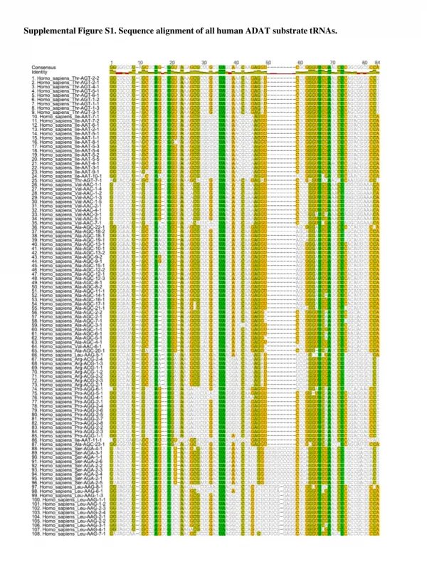
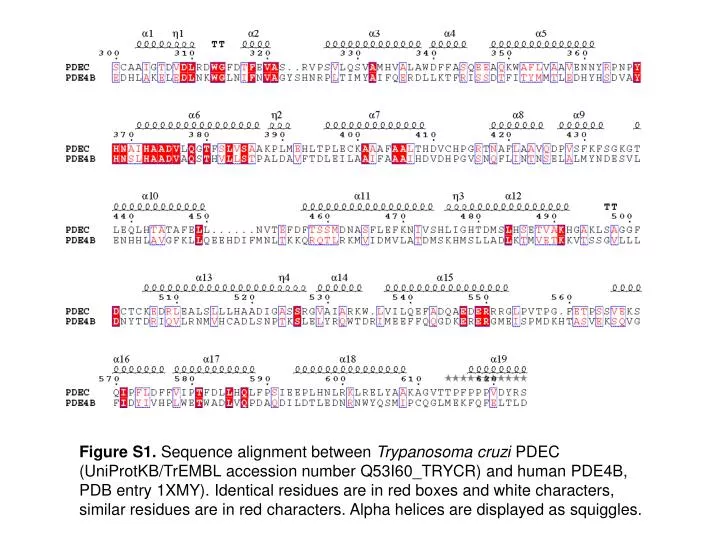
Figure S1. Sequence alignment between Trypanosoma cruzi PDEC (UniProtKB/TrEMBL accession number Q53I60_TRYCR) and human PDE4B, PDB entry 1XMY). Identical residues are in red boxes and white characters, similar residues are in red characters. Alpha helices are displayed as squiggles. A.

E N D
Figure S1. Sequence alignment between Trypanosoma cruzi PDEC (UniProtKB/TrEMBLaccession number Q53I60_TRYCR) and human PDE4B, PDB entry 1XMY). Identical residues are in red boxes and white characters, similar residues are in red characters. Alpha helices are displayed as squiggles.
A B Figure S2. A.The overlay of human PDE4B crystal structure (green ribbons) and Trypanosoma cruzi PDEC homology model (purple ribbons). B. The docking conformations of identified compounds around the active site of PDEC.