Download
1 / 79
860 likes | 1.2k Vues
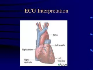
ECG Interpretation Part 2. Carolyn M. Malone RN,MSN,FNP-C. Review of Rhythms. Atrial Arrhythmia. Premature atria contraction or PAC impulse occurs earlier than predicted in the cycle causing the p-wave to look different. Usually does not require any treatment unless the patient is symptomatic.

E N D
ECG InterpretationPart 2 Carolyn M. Malone RN,MSN,FNP-C
Atrial Arrhythmia • Premature atria contraction or PAC impulse occurs earlier than predicted in the cycle causing the p-wave to look different. Usually does not require any treatment unless the patient is symptomatic
Atrial Flutter Characterized by classis saw-toothed waves instead of classis p-wave. If there is a rapid ventricular response this rhythm is grouped as Narrow Complex Tachycardia.
Narrow Complex Tachycardia Treatment • This term is assigned to tachyarrhythmias with a narrow QRS typically seen with atrial fibrillation or atrial flutter with rapid ventricular responses or A-Fir RVR • Assess ABC’S → apply oxygen →place patient on monitor → obtain blood pressure and oximetry → obtain IV access Is the QRS narrow (0.12 seconds)?
Narrow Complex Tachycardia Treatment • Is the rhythm regular? If yes attempt vagal maneuvers → administer Adenosine (Adenocard) 6mg rapid IV push prefer IV site at a/c level If not effective may repeat at 12 mg IV rapid push. Adenosine slows conduction time through the AV node and reentrant pathways restoring NSR. If the rhythm does convert continue to monitor the patient. If it does not convert consider other medications.
Narrow Complex Tachycardia Treatment • Diltiazem (Cardizem) inhibits calcium from entering the slow channels during depolarization producing a relaxation of vascular smooth muscle and coronary vasodilatation and increases myocardial oxygen delivery in patients with vasospastic angina. • Dose is typically 0.25mg/kg typically 20mg bolus over 2 minutes may repeat at 0.35mg/kg, usually 25mg bolus. If patient rhythm is not resolve pt will require drip typically at 5-10mg/hr
Narrow Complex Tachycardia Treatment • Verapamil (Calan) Inhibits calcium from entering the slow channels during depolarization, produces relaxation of coronary smooth muscle and coronary vasodilation, increases myocardial oxygen delivery, slows automaticity and conduction of AV node. • Dose 2.5-5mg SIVP over 2 minutes second dose at 5-10 mg 15-30 minutes after the first dose.
Narrow Complex Tachycardia Treatment • Betablockers Esmolol (Brevibloc). Competitively blocks response to β1 adrenergic stimulation with little or no effect on β2 receptors except at high doses. • Dose is 500mcg/kg/min loading dose followed by a 50mcg/kg/min for 4 minutes and continue to increase at 50mcg/kg/min to achieve rate control.
Intro to AV Blocks • It is the AV Node that is diseased in atrioventricular blocks. The SA node usually functions normally. • Since the SA node is not affected in AV blocks, the P waves occur at regular rhythm.
AV Blocks • Since the PR interval reflects the impulse traveling through the AV node this measurement is one of the criteria used to determine the type of AV block that is present. • A prolonged PR interval signifies first-degree AV block.
AV Blocks • There are three levels of AV block: first-degree, second-degree, and third-degree AV Block. • The number and pattern of impulses conducted through the AV node determines the presence and degree of AV block.
First-Degree AV Block • In first-degree AV Block, there is a 1:1 ratio between P waves and QRS complexes. • All of the impulses are conducted to the ventricles, but are delayed in the AV node. • The delay results in a prolonged PR interval. • The occurrence of first-degree AV block will be noted in addition to your interpretation of the patient’s basic rhythm. There is no tx.
ECG Criteria: First-Degree AV Block • P waves: similar, 1:1 with QRS • Rhythm: Atrial – regular Ventricular - regular • Rate: Usually 60-100/minute • PR: greater than 0.20 second (prolonged) • QRS: 0.04-0.10 second (normal) • QT: 0.32-0.44 second (normal)
Possible Causes of First-Degree AV Block • Conduction system disease • Digitalis administration • Antiarrhythmics, such as quinidine and Amiodarone • Acute MI
Treatment First-Degree AV Block • Treat underlying cause • O2, Atropine for symptomatic bradycardia
Second Degree AV Block • There are 2 type of second-degree AV block: • Mobitz I (also called Type I or Wenckebach) • Mobitz II (also called Type II) • The PR interval abnormality differentiate the two rhythms. • Mobitz II is a more serious dysrhythmia because the causes more often result in permanent changes. Mobitz II is also more likely to develop into complete or third-degree AV block.
Mobitz I - Wenckebach • With Mobitz I, the PR interval gradually lengthens until an impulse is not conducted. You will see a P wave without a corresponding QRS complex. The R-to-R shortens until a QRS is dropped. • It is as though the AV node is warning that soon an impulse won’t get through. • The P wave may be buried in another wave.
ECG Criteria Mobitz Type I Second Degree AV Block • P waves: similar – more P waves than QRS’s • Rhythm: Atrial – regular Ventricular - irregular • Rate: Atrial: usually 60-100/minute Ventricular – depends on the number of blocked impulses; will be less than atrial • PR: gradually increases until there is a P wave with no QRS; it is a cyclic pattern. • QRS: 0.04-0.10 second (normal) • QT: 0.32-0.44 second (normal)
Possible Causes for Mobitz I • Increased vagal stimulation (such as vomiting or straining) • Post myocardial infarction (usually inferior wall of left ventricle) • Medications (such as digitalis, beta-blockers, Quinidine, Procainamide)
Treatment - Mobitz Type I • No treatment is usually needed for Mobitz I as the rhythm is well-tolerated. Assess ABC’S apply oxygen, place the patient on the monitor and oximetry, establish IV access obtain blood pressure monitor for s&s of instability i.e chest pain, SOB, hypotension, chest pain, signs of shock, continue to monitor AV block, such as Mobitz II or third degree.
Second-Degree AV Block Mobitz II • With Mobitz II, the PR interval is constant, but some impulses are not conducted. There are P waves without QRS complexes. Unlike Mobitz I there is no warning that some of the impulses will be blocked.
ECG Criteria: Mobitz II Second-Degree AV Block • P waves: similar, more P waves that QRS • Rhythm: Atrial – regular Ventricular - irregular • Rate: Atrial – usually 60-100/minute Ventricular – depends on the number of blocked impulses; will be less that atrial rate • PR: constant for conducted beats • QRS: may be normal but often > 0.10 second • QT: 0.32-0.44 second (normal)
Possible Causes for Mobitz II • Acute Myocardial Infarction – usually anterior wall of left ventricle • Hypoxia • Chronic hypertension • Ischemic heart failure • Symptoms depend on frequency of dropped beats/ventricular rate • The causes of Mobitz II are likely due to irreversible damage to the conduction system
Treatment • The physician should be notified as soon as possible when this rhythm develops. Mobitz II is often associated with a poor prognosis since development of complete AV block should be anticipated. A temporary pacemaker may be inserted prophylactically/permanently • Assess ABC’S provide oxygen, place on monitor establish rhythm obtain blood pressure, oximetry, obtain IV access • Atropine dose is 0.5mg may be repeated up to a total of 3mg prepare to transcutaneous pace • Epinephrine 2-10mcg/min Dopamine 2-10mcg/kg/min vasoactive drips
Third-Degree AV Block • Complete (or third-degree) AV block indicated absence of conduction between the atria and ventricles. • The block can be in the AV node or in the bundle of His. The atria are paced by the SA Node and an independent pacemaker generates impulses for the ventricles. Thus, the atrial and ventricular contractions are not synchronized.
Third degree AV Block • The ECG rhythm is characterized by separate, but regular, atrial and ventricular rhythms and rates. Because of the different rates and because there are two separate pacemakers, there is no correlation between the P waves and the QRS complexes. As a results, there is no true PR interval.
ECG Criteria: Third-Degree AV Block • P waves: similar – more P waves than QRS’s • Rhythm: Atrial – regular Ventricular - regular • Rate: Atrial – usually 60-100/min Ventricular – usually less than 40/min; it can be up to 60/min if the block is high in the AV node • PR: varies – no true PR interval since atria and ventricles conducting independently • QRS: Greater than 0.10 second • QT: may be normal or prolonged
Possible Causes for Third-Degree AV Block • Extensive myocardial infarction (usually anterior wall of the left ventricle) • Acidosis • Hypoxia • Hyperkalemia • Digitalis toxicity • Extensive ventricular conduction system disease
Treatment – Third-Degree Block • The MD should be notified immediately when third-degree heart block develops. The patient may not tolerate a sudden decrease in cardiac output associated with the slow rate. • Because the independent ventricular pacemaker may be unreliable and may result in periods of asystole, third-degree heart block may become a lethal dysrhythmias. • A temporary transcutaneous or transvenous pacemaker is usually inserted. A permanent cardiac pacemaker is often necessary.
Treatment – Third-Degree Block • Assess ABC’S → administer oxygen → obtain blood pressure, place on ECG monitor oximetry → establish IV access → assess perfusion status i.e. chest pain, mental status, hypotension, signs of shock → prepare for transcutaneous pacing → consider Atropine at 0.5mg IV may repeat for a total dose of 3mg → consider Epinephrine drip at 2-10mcg/min or Dopamine 2-10mcg/kg/min while waiting for pacer or if pacing is ineffective.
Modes of Pacing Synchronous (demand) pacing 1. Temporary pacing most common 2. Pacemaker's sensitivity set to client's beats 3. If intrinsic rate is rate set, the pacemaker is inhibited from firing 4. If intrinsic rate is rate set, pacemaker fires to stimulate depolarization Asynchronous (fixed rate) 1. Used for asystole or profound bradycardia 2. Fires at a fixed rate regardless of client's intrinsic rhythm 3. Complications: pacemaker competition, R-on-T phenomenon 34-24
Noninvasive Temporary Pacing (NTP) Two large patch electrodes attached to an external pulse generator Electrical pulses transmitted transcutaneously to stimulate depolarization Used as an emergency measure or prophylactically Complications: discomfort from cutaneous/muscle stimulation, skin irritation and diaphoresis from patches Loss of capture Inappropriate pacing 34-25
Figure 34-20AEquipment and electrode placement for external pacing
Figure 34-20BEquipment and electrode placement for external pacing
Invasive Temporary Pacing Transvenous pacing Epicardial pacing Complications: 1. Infection/hematoma 2. Ectopic complexes 3. Loss of capture 4. Under-sensing/pacemaker competition/over-sensing 5. Electromagnetic interference 6. Stimulation of chest wall or diaphragm 34-26