Download
1 / 19
190 likes | 275 Vues
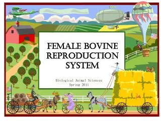
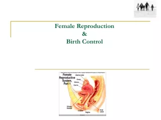
Female Bovine Reproduction System. By: Natalia Bahena. Reproductive System. http://cvm.msu.edu/courses/AP/bessie/cowreproorgans3.jpg. We are going to look into the function of the hypothalamus and the pituitary gland.

E N D
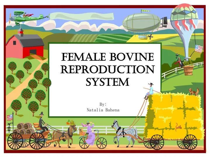
Female BovineReproduction System By: Natalia Bahena
Reproductive System http://cvm.msu.edu/courses/AP/bessie/cowreproorgans3.jpg
We are going to look into the function of the hypothalamus and the pituitary gland.
Two essential organs of reproduction are located within the head of the animal. The hypothalamus controls: Body temperature, and the drive to eat and drink are just a few functions. It sends and receives neural signals through the nervous system and hormonal messages through the endocrine system. The pituitary gland, sits at the base of the brain. The pituitary is divided into two regions: the anterior and posterior pituitaries.
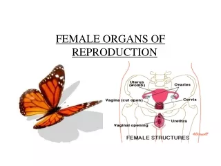
http://www.ca.uky.edu/agripedia/Classes/ASC106/media/FEMALE.GIFhttp://www.ca.uky.edu/agripedia/Classes/ASC106/media/FEMALE.GIF • The female reproductive organs consist of the ovary, uterus, cervix, vagina and vulva. Female reproductive tracts of various farm animals are similar to the cow, but differ primarily in the shape of the uterus and cervix.
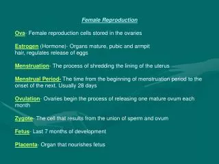
The ovary, is responsible for two basic functions: Production of the female egg or ovum. Production of two primary reproductive hormones, estrogen and progesterone. Ovaries
http://www.ansi.okstate.edu/course/3443/study/AnatomyFemale/bovine/sld015.htmhttp://www.ansi.okstate.edu/course/3443/study/AnatomyFemale/bovine/sld015.htm The oviduct begins as a funnel-shaped tube that engulfs the ovary. When ovulation occurs, the ovum is picked up by the infundibulum and channeled into the oviduct (also known as the Fallopian tube), where fertilization takes place if sperm are present.
The uterus of the cow is bipartile, while the uterine horns are relatively long and well developed. The fertilized embryo moves from the oviduct into the uterine horn, where fetal development begins. The fetus grows within a layer of membranes called the placenta, where it is nourished. Uterus ianrpubs.unl.edu/ beef/g537.htm
Cervix The cervix has annular rings. It has thick walls that allow a passageway for sperm at mating and expulsion of the fetus at the time of birth. During pregnancy, the cervix is filled with a thick mucus secretion known as the cervical plug, which protects the uterus from infections entering from the vagina. http://www.ansi.okstate.edu/course/3443/study/Notes/female/cervix.jpg
The vagina serves as a receptacle for the male's penis. • In the cow, the semen is deposited in the vagina near the cervix during natural mating with the bull. • When artificial insemination is used, an insemination instrument is threaded through the vagina and cervix and semen is deposited at the uterine side of the cervix. • The external opening of the vagina is called the vulva. http://www.ansi.okstate.edu/course/3443/study/AnatomyFemale/bovine/img024.JPG
Estrous Cycle • The ovarian changes during a typical 21-deay estrous cycle in which pregnancy does not occur. • The development and regression of the corpus luteum and of the follicles are continuous processes http://www.cahe.nmsu.edu/pubs/_b/b-212.pdf
. This cycle of egg development in cattle is called the estrous cycle. The cow is a non seasonal polyestrous species. Which means a cow can have multiple estrous cycles throughout the year. Two prominent structures are present within the ovary, the follicle and corpora lutea. http://www.aces.edu/pubs/docs/A/ANR-1027/
http://207.62.207.35/vet02/vett2/vett2photos/lecture/module23/photo06.gifhttp://207.62.207.35/vet02/vett2/vett2photos/lecture/module23/photo06.gif Estrus heat is not always accompanied by ovulation, nor ovulation by estrus. Heat without ovulation will not result in pregnancy even though the female is bred.
Hormones • Estrogen- • Prepares the pre-pubertal heifer and post-partum cow for cyclic sexual activity. • Progesterone, secreted by the corpora lutea, suppresses the further development of follicles and the secretion of estrogen. High levels of progesterone and low levels of estrogen prevent a cow from coming into heat. Progesterone is necessary for preparing the uterus to receive the fertilized egg and maintains the proper uterine environment for continuation of pregnancy. • Follicle stimulating hormone (FSH) and luteninizing hormone (LH) are secreted & travel through the blood to the ovary. • FSH and LH are mediated by gonadotropic releasing hormone (GnRH) coming from the hypothalamus to signal their release from the pituitary. • FSH stimulates the growth, development and function of the follicle, while LH cause the follicle to rupture during ovulation and causes the subsequent development of the corpus luteum. http://www.mothercow.org/bull/barnyard/hormones.jpg
Placenta • Classification is based on: • The gross shape of the placenta and the distribution of contact sites between fetal membranes and endometrium. • The number of layers of tissue between maternal and fetal vascular systems. http://arbl.cvmbs.colostate.edu/hbooks/pathphys/reprod/placenta/plac_types.jpg
http://arbl.cvmbs.colostate.edu/hbooks/pathphys/reprod/placenta/structure.htmlhttp://arbl.cvmbs.colostate.edu/hbooks/pathphys/reprod/placenta/structure.html
The three potential maternal layers in a placenta are: • Endothelium lining endometrial blood vessels. • Connective tissue of the endometrium. • Endometrial epithelial cells. http://arbl.cvmbs.colostate.edu/hbooks/pathphys/reprod/placenta/structure.html
A cow has a cotyledonary placenta. Cotyledonary: Multiple, discrete areas of attachment called cotyledons are formed by interaction of patches of allantochorion with endometrium. The fetal portions of this type of placenta are called cotyledons, the maternal contact sites (caruncles), and the cotyledon-caruncle complex a placentome. This type of placentation is observed in ruminants. http://www.uoguelph.ca/zoology/devobio/splab9/sld012.gif
http://arbl.cvmbs.colostate.edu/hbooks/pathphys/reprod/placenta/structure.htmlhttp://arbl.cvmbs.colostate.edu/hbooks/pathphys/reprod/placenta/structure.html