Download

1 / 39
390 likes | 513 Vues
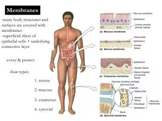
Membranes. -many body structures and surfaces are covered with membranes -superficial sheet of epithelial cells + underlying connective layer. -cover & protect. -four types:. 1. serous. 2. mucous. 3. cutaneous. 4. synovial. 1. Serous membranes.

E N D
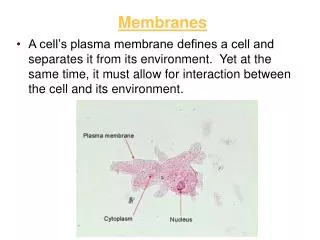
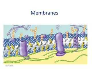
Membranes -many body structures and surfaces are covered with membranes -superficial sheet of epithelial cells + underlying connective layer -cover & protect -four types: 1. serous 2. mucous 3. cutaneous 4. synovial
1. Serous membranes -line the subdivisions of the abdominopelvic cavity and thoracic cavity -covers, protects and moistens/lubricates -comprised of an epithelial layer (simple squamous epithelium) called a mesothelium + underlying loose areolar connective tissue -the mesothelium secretes a watery fluid = serous fluid (separates and lubricates the movement of organs) -divided into two separate layers: 1)outer parietal layer - lines the cavity 2) inner visceral layer - covers organs -serous membrane lining the pleural cavity (lungs) = pleura - serous membrane lining the pericardial cavity (heart) = pericardium - serous membrane lining the peritoneal cavity (abdomen) = peritoneum
2. Mucous membranes -line cavities that directly communicate with the exterior environment e.g. respiratory, urinary, reproductive, digestive -covers, protects and moistens/lubricates -epithelial layer (simple squamous, simple cuboidal, simple columnar) is kept moist through production of mucus by glands, other glandular secretions or exposure to fluids (e.g. urine) -in areas of physical stress = stratified epithelial tissue rather than simple -connective tissue layer is loose areolar tissue = lamina propria --supports embedded blood vessels and nerves
3. Synovial membranes -extensive areas of areolar connective tissue covered by incomplete layers of simple squamous or cuboidal epithelial cells -lines & lubricates the synovial joint cavity - to permit easy movement of bones -the epithelium differs from others: 1) there is no basal lamina 2) incomplete cellular layer - gaps between cells 3) derived from macrophages and from the surrounding connective tissue -some cells within this membrane are phagocytic to remove pathogens -others are secretory - secrete a watery synovial fluid for lubrication
4. Cutaneous membrane (skin) -covers the surface of the body -epithelial layer (keratinized stratified squamous) -underlying areolar tissue reinforced with dense connective tissue
Integumentary System (Skin) • skin covers the entire body surface • -including the anterior surface of the eye! • -covers ~ 22 square feet • -about 16% of total body weight • skin turns in at the mouth, nasal cavity, anus and urethral and vaginal • openings – meets the mucous membranes lining these cavities • comprised of all four tissues: • 1. epithelium – lines the surface • 2. connective – provides strength & resiliency • 3. muscle – smooth muscle controls blood vessel diameter • and controls movement of hairs • 4. nervous – provides sensation and controls SM
functions: 1. physical protection: protection from microbes, abrasion, heat 2. chemical protection – keratin - dryness of the epidermis; salt of sweat 3. regulation of water exchange: by sweating 4. regulation of body temperature: thermoregulation -by sweating & adjusting blood flow through the dermis 5. excretion of wastes -by sweating 6. nutrition – synthesis of vitamin D precursor -activated in skin, converted to calcitrol in liver 7. sensation: touch, pressure, vibration, pain & thermal 8. immune defense: Langerhans cells of the epidermis
-two major components: 1. cutaneous membrane = skin (epidermis, dermis) 2. accessory structures = hair, nails, exocrine glands
Epidermis -stratified squamous epithelium - 5 layers maximum -four types of cells: 1. keratinocytes – make up the majority of the epidermis -epithelial cells that synthesize the protein keratin 2. melanocytes – cells for the synthesis of the light absorbing pigment melanin 3. Merkel cells – neurons that detect pressure 4. Langerhans cells – immune responses
Epidermis: layers - stratum germinativum: -inner most/deepest layer of the epidermis -also called stratum basale because it attaches firmly to the basal lamina or basement membrane found between the epithelium and connective tissues of the dermis - contain basal stem cells that differentiate into the keratinocytes and melanocytes of the epidermis -Merkel cells are found in hairless regions -pressure and touch receptors of the skin
Epidermis: layers - stratum spinosum: -called the “spiny layer” because of histological appearance following chemical treatment -keratinocytes of the stratum basale migrate into this layer -several layers thick -keratinocyotes interconnected by bundles of protein filaments called tonofibrils – connect neighbouring keratinocytes together -act as cross braces providing strength -cells can divide - division of cells within this layer increases thickness -melanocytes are common -Langerhans cells also found – in the more superficial layers of this layer -initiate immune responses to pathogens and to cancer
Epidermis: layers -stratum granulosum: -made up of keratinocytes migrating up from the stratum spinosum -cells synthesize large quantities of proteins (including keratin) – cytoplasm appears granular -the granules = keratohyalin granules -these granules surround the keratin filaments as they develop -as keratin is made – keratinocytes become thinner and flatter -the cells then die and dehydrate -creates layers of interlocking keratin “sandwiches”
Epidermis: layers stratum lucidum: -covers the Str. Gran. -flattened, densely packed cells filled with keratin -have a glassy appearance because they do not stain well -present only in the skin of fingertips, palms & soles ONLY FOUND IN THE PALMS OF THE HAND SOLES OF THE FEET
Epidermis: layers - stratum corneum: -cornu = horn -makes up surface of both thick and thin skin -15-30 layers of flattened, dead, interlocking cells -large amounts of keratin are present – the tissue is said to be “cornified” -covered in secretions from dermal glands to help moisturize the outer layer -but keratin makes this layer water-resistant - very dry – prevents growth of microorganisms -most of this layer is also hydrophobic -penetration is promoted by attachment to a lipid or dissolution in a lipid-based solution -transdermal drug patches – drugs are in oils or lipid-soluble carriers - moisturizing lotions – only penetrate few first layers of corneum -takes 15-30 days to move from germinativum to corneum -cells will remain in corneum for an additional 2 weeks before being shed
Dermis -two major components: 1. papillary layer 2. reticular layer Papillary Layer -about 1/5th thickness of dermis -loose areolar connective tissue + elastic fibers -contains capillaries for blood supply & sensory nerve endings -dermal projections into the epidermis = papillae (papilla – “nipple-shaped mound) -some papillae contain Meissner’s corpusclesfor touch -also free nerve endings – project into the epidermis - sensations of pain, warmth, itching Reticular Layer (“little net”) -dense irregular connective – interwoven collagen bundles plus elastic fibers -contains blood vessels, nerves, hair follicles, sweat glands and sebaceous/oil glands -also contains lamellated corpuscles (Pacinian corpuscles) that detect deep touch and pressure
Fingerprints: epidermal ridges -formed from the stratum germinativum -extends down into the dermis -formed by the connections between dermal papillae and the epithelium -the contours of the skin follow these ridge patterns = fingerprints -function to increase the SA of the skin and increase friction
Skin colour • dermal blood supply: • - hemoglobin bound to O2 – bright red in color • -gives pinkish cast to skin • -when hemoglobin lacks O2 – bluish colour • -this bluish skin colour = “cyanosis” • -the thin skin of the lips and transparency of the nail enables us to see the blood in the peripheral circulation = red lips and pink nails • -dermal blood supply comes from the larger blood vessels found in the subcutaneous layer (located under the dermis) • -these larger vessels branch to form a cutaneous plexus (plexus = network) - this supplies the reticular layer • vessels continue up and branch further into the papillary plexus - found in the dermal papillae • -blood is drained out of the papillae by tiny veins = venules • -these drain into the larger veins of the dermis -> which then drain into the SQ layer
Skin colour 2. pigmentation – two pigments: carotene and melanin -carotene = orange, yellow colour -derived from vitamin A -can be converted back to vitamin A in the skin - required for epithelial maintenance & the synthesis of visual pigment (rhodopsin) -carotene accumulates in keratinocytes -yellow color is very evident in the stratum corneum
-melanin = dark brown, black colour -synthesized from the amino acid tyrosine -melanin absorbs UV light and prevents damage to the keratinocytes of skin -melanin production stimulated by UV light -produced by the melanocytes of the epithelium -forms in intracellular vesicles = melanosomes -melanosomes are secreted out of the cells = melanin is transferred into keratinocytes -Caucasians: transfer occurs to keratinocytes only in the Str. germ. and spinosum-Blacks: larger melanosomes -transfer also occurs in the Str. granulosum -darker pigmentation results -more active melanocytes – NOT more in number!
Wrinkles: reduction in the thickness of the dermis -loss of collagen in the dermal reticular layer -loss in dermal flexibility = wrinkles and sagging SOME FUN STUFF TO KNOW ABOUT SKIN Scars: from greek word schara (place of fire) -damaged dermis is replaced with tissue of inferior quality and rich in collagen -scars do not have sweat or oil glands and do not have hair -redness of the scar is due to inflammation and is not permanent - two common types: 1. hypertropic (red and raised, do not grow beyond boundaries of original wound 2. keloid – permanently growing scars – can lead to benign tumors - more common in darker skin, common on chest and shoulders Stretch marks: extensive and quick distortion of the dermis damages it -no recoil of skin after stretching -leads to breaking of elastic and collagen fibers in the dermis - replaced with new, poorly organized collagen Retin-A (tretinoin) : increases blood flow to the dermis -promotes dermal repair -decreases wrinkles and stretch marks
Subcutaneous layer -also referred to as the hypodermis or superficial fascia -connects the skin to underlying muscles or other organs -made up of: 1. loose connective tissue 2. adipose tissue - “baby fat” - also contains elastic fibers for flexibility -fat content helps reduce heat loss -fat distribution changes with age and gender: -males – neck, upper arms, abdomen and lower back -females – breasts, abdomen, buttocks, hips and thighs -contains large arteries and veins – supply the dermal plexuses with blood -the superficial layers of the hypdermis are the sites for drug injections - hypodermic
Accessory Structures Hair follicles Sweat glands Sebaceous/Oil glands Nails
Hair & Hair follicles • over all epidermal surfaces except soles of feet, palms of hand, sides of fingers and toes and portions of external genitalia • approx. 5 million hairs on the body • formed in hair follicles • Comprised of three major regions: • 1. Hair bulb – contains the living, hair • papilla • 2. Hair root – site of connection with • arrector pili (smooth muscle) and a • sebaceous gland • 3. Hair shaft – portion of the hair above the sebaceous gland • -portion of it is exposed above the skin
Hair structure -hair papilla – found at the base of the hair within the hair bulb -contains the stem cells of the hair = hair matrix -also contains capillaries and sensory nerves for touch -epithelial tissue surrounding the matrix make up the hair bulb -hair root: projects up from the hair bulb -surrounded and protected by a hair follicle -site of attachment of arrector pili smooth muscle (pulls hair upright for better sensation) -continues above the sebaceous gland as the hair shaft – also exposed above the surface of the skin
Hair Production -hair production = specialized keratinization -hair is produced from the hair matrix – epithelial layer similar to stratum basale (contains living cells of the hair - many are stem cells) -keratinocytes differentiate within the matrix – cells immediately produce the keratin of hair -keratin forms into an outer cortex and inner medulla -cortex – hard keratin - stiffness -medulla – soft keratin – flexible -single layer of dead cells encloses the cortex of the hair = cuticle -within the dermis – hair is enclosed in a protective follicle (made also by the hair matrix
Follicle -found in the dermis surrounding the hair -often rooted in SQ layer -wall is comprised of four layers: 1. internal root sheath: surrounds the root & deeper portion of shaft -produced by the cells of the hair matrix 2. external root sheath: runs continuously from the hair bulb to the skin surface to enclose the hair 3. glassy membrane: thickened basement membrane between external root sheath and the connective tissue sheath 4. connective tissue sheath
Exocrine Glands • secretions are discharged out onto the surface of the epithelium that lines • body cavities or out onto the skin • many exocrine glands secrete to the exterior via tubes called ducts • exocrine secretions: • 1. perspiration • 2. digestive enzymes • 3. milk • 4. mucous • 5. oil
Exocrine glands • you can classify exocrine glands many ways • one way – by the consistency of what they secrete • e.g. serous • another way – by their structure • e.g. multicellular • last way – by the mode of secretion • e.g. holocrine
exocrine gland types – consistency of secretion: • 1. serous - watery fluid that contains enzymes • e.g. saliva – parotid salivary gland • 2. mucous - glycoproteins called mucins that absorb water to form a • slippery mucus • e.g. sublingual salivary gland • 3. mixed - more than one type of gland cell • -produces different types of secretions - mucus and serous • e.g. submandibular salivary gland
exocrine gland structure: • Unicellular are single-celled glands • e.g. goblet cells • Multicellular glands • -two characteristics to classify: • shape of the secretory portion • branching pattern of the duct • -simplest multi-cellular gland is a secretory sheet • secrete into a compartment • e.g. gastric epithelium
Modes of Secretion: Exocrine glands • merocrine: • -contents are released through exocytosis
2. apocrine: loss of cytoplasm from the apical portion of the cell together with secretory product e.g. milk secretion
3. holocrine: results in death of gland cell -entire cell fills with secretory product and then bursts e.g. sebaceous glands associated with hair follicles
Skin glands 1. Sweat/Sudoriferous – of the serous type 2. Oil/Sebaceous – of the mucus type 3. Wax/ Ceruminous Sebaceous glands/Holocrine glands -”sebace” = greasy -secreting portion is within the dermis -most open onto hair follicles -glands located at the lips, glans penis, labia minora and eyelids - open directly to the skin surface -absent on palms and soles -large in size and numbers on breast, face, neck and upper chest
sebaceous glands secrete an oily substance = sebum • acne = inflammation of a sebaceous gland due to the presence of bacteria • may cause a cyst or sac of connective tissue to form which destroys epithelial cells • known as cystic acne
Sudoriferous Glands • 3 to 4 million glands in the body • sudori = sweat, ferous = bearing • released by exocytosis into hair follicles or onto the skin surface • two main types: • 1. Merocrine sweat glands (eccrine) • simple, coiled tubular glands - serous • secretion through exocytosis • found throughout the skin, PLUS margins of lips, nail beds, glans penis and clitoris • most numerous in forehead, palms and soles • secretory portion is located in the reticular layer of dermis • ends as a pore in the skin • main function is to regulate body temperature through evaporation • also functions in waste elimination • thin, watery perspiration - about 600 ml per day • -water, sodium, chloride, urea, uric acid, ammonia, glucose, amino acids • -perspiration can be: a. insensible - evaporates before being perceived • b. sensible - larger amounts, can be seen and felt
Apocrine sweat glands • makes odorous secretion - secretions are slightly more viscous than merocrine • simple, coiled tubular glands – secretory portion in hypodermis • skin of axilla (armpit), groin, areolae, bearded region of face in males • released by a portion of the cell breaking off and disintegrating • secretory portion located in SQ layer - opens onto a hair follicle • same components as sweat + lipids and proteins • do not function until after puberty Sudoriferous gland – merocrine/apocrine
Mammary glands • Large, complex apocrine sweat glands • Ceruminous glands • modified sweat glands • in ear canal • secretory portion is in the SQ layer, deep to sebaceous glands • secrete either directly into the ear canal or into the ducts of • the sebaceous glands • produce waxy cerumin - together with the hairs of the canal • provides protection
Nail Anatomy • Nail body:visible • free edge projects beyond digit • pink due to blood flow • proximal end is white crescent • called a lunula-thickened str. basale • hyponychium: secures the nail to the finger • Nail root: • -production of nail • -buried in a fold of skin • -deep to the root = nail matrix • Cuticle (eponychium) • -fold of stratum corneum • -adheres nail to the lateral borders of the nail wall