Download
1 / 1
10 likes | 385 Vues
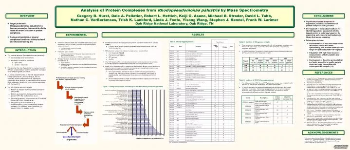
Typical Results Table 1. Affinity-tagged proteins Gene # Name Description Unique peptides % sequence coverage # trials RPA0176 atpD ATP synthase beta subunit 35 79 7 RPA0190 sucD succinyl-CoA synthetase alpha-subunit 18 60 1 RPA0191 sucC succinyl-coA synthetase beta chain

E N D
Typical Results Table 1. Affinity-tagged proteins Gene # Name Description Unique peptides % sequence coverage # trials RPA0176 atpD ATP synthase beta subunit 35 79 7 RPA0190 sucD succinyl-CoA synthetase alpha-subunit 18 60 1 RPA0191 sucC succinyl-coA synthetase beta chain 16 60 1 RPA0192 mdh malate dehydrogenase 41 76 1 RPA0245 ffh/ftsY signal recognition particle 1 1.4 1 RPA0962 hupS uptake hydrogenase small subunit 1 5 2 RPA0963 hupL uptake hydrogenase large subunit 45 67 6 RPA1141 groES 1 small subunit of GroESL molecular chaperone 9 65 3 RPA1548 puhA H subunit of photosynthetic reaction center ND ND 2 RPA1980 cbbZ phosphoglycolate phosphatase ND ND 1 RPA1981 cbbI, rpiA, ppi ribose 5-phosphate isomerase 28 82 2 RPA2164 groEL 2 large subunit of GroESL molecular chaperone 62 72 9 RPA2165 groES 2 small subunit of GroESL molecular chaperone 6 41 4 RPA2336 RPA2336 unknown protein 26 73 3 RPA2339 possible iron response transcription regulator ND ND 1 RPA2405 draT NAD+ ADP-ribosyltransferase ND ND 1 Figure 1. Background proteins observed by LC-MS-MS in Affinity Isolation Experiments RPA2406 draG dinitrogenase reductase activating ND ND 1 RPA2462 RPA2462 conserved unknown protein 2 23 1 RPA2867 pyruvate dehydrogenase E1 alpha subunit ND ND 1 putative H+-transporting ATP synthase alpha chain., RPA0178 30S ribosomal protein S2, RPA2922 RPA2967 glnA glutamine synthetase I 36 56 1 50S ribosomal protein L2, RPA3247 two-component transcriptional regulator, winged helix family, RPA1930 RPA2969 RPA2969 unknown protein 8 40 2 unknown protein, RPA2552 RPA3147 clpA endopeptidase Clp: ATP-binding chain A ND ND 1 peroxiredoxin-like protein, RPA4268 conserved hypothetical protein, RPA2050 RPA3226 rpoA DNA-directed RNA polymerase alpha subunit 37 73 2 elongation factor Tu, RPA3252 RPA3247 rplB 50S ribosomal protein L2 9 42 1 DUF156, RPA1661 putative H+-transporting ATP synthase beta chain., RPA0176 RPA3248 rplW 50S ribosomal protein L23 13 72 1 UDP-N-acetylmuramate-alanine ligase, RPA3529 nitrogen regulatory protein PTSI(NTR), RPA0605 RPA3252 tufA, EF-Tu elongation factor Tu ND ND 2 phosphomethylpyrimidine kinase (hmp-phosphate kinase), RPA3971 RPA3253 fusA, EF-G elongation factor G 62 63 2 ribosomal protein S5, RPA3233 CBS domain, RPA1220 RPA3618 AAA ATPase ND ND 1 DUF88, RPA2691 possible DNA-binding protein hu-alpha (NS2) (HU-2), RPA2953 RPA3834 idh NADP-dependent isocitrate dehydrogenase 20 43 2 possible outer membrane lipoprotein GNA33, RPA0304 RPA3876 fumA fumarate hydratase, class I 45 41 2 possible dehydrogenase, RPA0422 transcriptional regulator, FUR family; probable FUR protein, RPA0450 RPA3878 conserved unknown protein 2 25 1 FeoA family, RPA4636 chaperonin GroEL1, cpn60, RPA1140 RPA4048 rfbF alpha-D-glucose-1-phosphate 8 30 1 conserved unknown protein, RPA1157 RPA4049 rfbG cdp-glucose 4,6-dehydratase 16 50 1 putative ribonuclease E, RPA2450 conserved unknown protein, RPA4191 RPA4050 unknown protein 18 62 1 dihydroxy-acid dehydratase, RPA3472 unknown protein, RPA3786 RPA4433 clpB Clp endopeptidase ATP-binding subunit 73 61 2 fructose-bisphosphate aldolase, RPA4642 RPA4464 soxC molybdopterin subunit sulfite oxidase 15 36 1 possible GTP cyclohydrolase II, riboflavin biosynthesis, RPA1093 chaperonin GroEL2, cpn60, RPA2164 RPA4465 soxB sulfite dehydrogenase 17 42 1 unknown protein, RPA1824 formyltetrahydrofolate deformylase, RPA4032 RPA4618 nifK nitrogenase beta subunit 19 47 1 possible CobW protein involved in cobalamin synthesis, RPA0861 RPA4619 nifD nitrogenase alpha subunit 38 63 1 conserved unknown protein, RPA4330 conserved unknown protein, RPA1653 RPA4620 nifH nitrogenase iron-protein 12 51 2 0 10 20 30 40 50 60 70 80 RPA4641 cbbM RuBisCo FormII 34 74 4 Frequency of appearance in MS measurement (%) RPA4644 cbbP phosphoribulokinase 33 76 2 ND: not detected by mass spectrometry Analysis of Protein Complexes from Rhodopseudomonas palustris by Mass SpectrometryGregory B. Hurst, Dale A. Pelletier, Robert L. Hettich, Keiji G. Asano, Michael B. Strader, David L. Tabb, Nathan C. VerBerkmoes, Trish K. Lankford, Linda J. Foote, Yisong Wang, Stephen J. Kennel, Frank W. LarimerOak Ridge National Laboratory, Oak Ridge, TN OVERVIEW CONCLUSIONS • Significant progress is reported in expression, isolation, and detection of protein complexes in R. palustris. • Development of more robust methods for identifying proteins associated with the tagged protein is underway, based on the expanding, well-characterized background library that we are amassing. • Future plans include: • Implementation of improved separations techniques, online with mass spectrometry, that provide wider dynamic range and potentially faster throughput • Integration with high-mass-accuracy measurements of both peptides and intact proteins. • Development of digestion protocols that are faster, amenable to smaller sample sizes, and more compatible with subsequent MS analysis. [10] • Target proteins in Rhodopseudomonas palustris have been expressed as fusions with affinity labels to enable isolation of protein complexes. • Component proteins of several complexes have been affinity-isolated and characterized by MS. RESULTS EXPERIMENTAL Table 2: Isolation of Nitrogenase complex • Three proteins in a nitrogenase complex (nifD, nifH, nifK) were each cloned with both His6 and V5 epitope tags, expressed under photoheterotrophic, nitrogen-fixing conditions, and isolated. • LC-MS-MS analysis of the isolate from each labeled component showed evidence for all three components of the nitrogenase complex. • Selected R. palustris genes were cloned with affinity tags [7] and expressed in both E. coli and R. palustris using modified pDEST vectors (Invitrogen). • Tag contains both His6 and V5 epitope • C-terminal position • Isolation of fusion proteins • Affinity purification with Ni-NTA agarose beads, followed by anti-V5 antibody agarose beads • Expression confirmed using 1-D PAGE and western blots. • “Shotgun” analysis: analysis by mass spectrometry without prior gel separation [8] • trypsin digestion • Reverse-phase HPLC separation online with electrospray/quadrupole ion trap MS-MS • Protein ID’s: Sequest [9] analysis of tandem mass spectral data using R. palustris database [1] . • Plasmids encoding 42 affinity-tagged fusion proteins have been inserted into R. palustris (Table 1). • Choice of target proteins guided by proteomics measurements (poster ThPT 382, VerBerkmoes et al.) • Multiple protein components of several complexes have been identified: • Iron nitrogenase (Table 2) • RNA polymerase (Table 3) • ATP synthase • GroESL • Over 400 “background” (i.e., unlabeled) proteins were commonly observed in numerous LC-MS-MS experiments. Figure 1 shows the most frequently detected of these. • Based on this expanding library of background measurements, we are currently developing criteria for determining whether detection of a given protein in a particular isolation/mass spectrometry experiment represents a valid “hit.” • One rough indication of a valid “hit” may be whether selected parameters for a detection (e.g. sequence coverage, number of unique peptides, total number of peptide spectra) significantly exceed those observed in the background for that protein. • As more measurements are made, the background library expands, and “hits” can be continually re-evaluated and refined. INTRODUCTION • The bacterial species Rhodopseudomonas palustris [1] • occurs widely in the environment • survives in a variety of conditions • light / dark • aerobic / anaerobic • This species thus has the potential to express markedly different complements of proteins and protein complexes under different growth conditions. • As part of a center funded by the U.S. Department of Energy Genomes To Life program [2,3], we are analyzing protein complexes from R. palustris by expressing target proteins as fusions with affinity tags to allow subsequent isolation of other proteins associated with the target [4]. • The MS analysis approach includes • Bottom-up analysis of affinity-isolated complexes (this poster) • Bottom-up proteomics of R. palustris proteins (poster ThPT 382, VerBerkmoes et al.) • Top-down analysis of intact proteins by FT-ICR-MS (poster MPU 390, Connelly et al.) • Integrated top-down and bottom-up characterization [5] of a conventionally-isolated complex (70S ribosome from R. palustris). [6] (poster ThPN 270, Strader et al.) REFERENCES 1. Larimer, F. W. et al., “Complete genome sequence of the metabolically versatile photosynthetic bacterium Rhodopseudomonas palustris.” Nature Biotech. 2004, 22, 55-61. 2. http://www.genomestolife.org 3. Buchanan, M. V. et al., “Genomes to Life ‘Center for Molecular and Cellular Systems:’ research program for identification and characterization of protein complexes.” OMICS2002, 6, 287-303. 4. (a) Gavin, A.-C. et al., "Functional organization of the yeast proteome by systematic analysis of protein complexes," Nature 2002, 415, 141-147. (b) Ho, Y. et al., "Systematic identification of protein complexes in Saccharomyces cerevisiae by mass spectrometry," Nature 2002, 415, 180-183. 5. Verberkmoes, N.C. et al., “Integrating “top-down” and “bottom-up” mass spectrometric approaches for proteomic analysis of Shewanella oneidensis,” J. Proteome Res.2002, 1, 239-252. 6. Strader, M.B., et al., “Analysis of the 70S ribosome from Rhodopseudomonas palustris using integrated top-down and bottom-up mass spectrometric approaches.” Sixth International Symposium on Mass Spectrometry in the Health and Life Sciences: Molecular and Cellular Proteomics, San Francisco, CA, Aug. 24-28, 2003. 7. Puig, O. et al., "The tandem affinity purification (TAP) method: a general procedure of protein complex purification," Methods 2001, 24, 218-229. 8. Link, A.J. et al., "Direct analysis of protein complexes using mass spectrometry," Nature Biotech. 1999, 17, 676-682. 9. Eng, J. K. et al., “An approach to correlate tandem mass spectral data of peptides with amino acid sequences in a protein database.” J. Am. Soc. Mass Spectrom.1994, 5, 976-89. 10. Russell, W. K. et al., “Proteolysis in Mixed Organic-Aqueous Solvent Systems: Applications for Peptide Mass Mapping Using Mass Spectrometry.” Anal. Chem.2001, 73, 2682-85. PCR amplification of target gene and cloning into modified expression vector Table 3: Isolation of RNA Polymerase complex • The alpha subunit of a DNA-directed RNA polymerase complex was cloned with both His6 and V5 epitope tags, expressed in R. palustris, and isolated. • LC-MS-MS analysis of the isolate showed evidence for the beta, beta’, and omega subunits of the complex. Additionally, two sigma factors for this polymerase were observed. These sigma factors are sub-stoichiometric, and associate with the polymerase under different conditions of growth or stress. Transfer expression plasmid into R. palustris Expression of affinity tagged proteins under various physiological conditions Capture protein complexes using affinity tag Characterize Protein Complex ACKNOWLEDGEMENTS Stoichiometries Functional assays Mass Spectrometry This research sponsored by the Office of Biological and Environmental Research, U.S. Department of Energy. Oak Ridge National Laboratory is managed by UT-Battelle, LLC, for the U.S. Department of Energy under Contract No. DE-AC05-00OR22725. ID proteins