Download
1 / 85
860 likes | 1.08k Vues
Aortic aneurysms and anesthesia. Moderator: Dr. Renu Presenters: Dr. Dipal Dr. Mridu. www.anaesthesia.co.in anaesthesia.co.in@gmail.com. Sub acute aortic dissection Expanding aortic aneurysm. Stable aortic aneurysm Coarctation of aorta Atherosclerotic disease.

E N D
Aortic aneurysms and anesthesia Moderator: Dr. Renu Presenters: Dr. Dipal Dr. Mridu www.anaesthesia.co.in anaesthesia.co.in@gmail.com
Sub acute aortic dissection • Expanding aortic aneurysm • Stable aortic aneurysm • Coarctation of aorta • Atherosclerotic disease • Bioprosthetic valve • Graft failure • Progression • Pseudo aneurysm
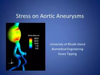
Aortic Aneurysm: • Definition: • Dilatation of aorta containing all the 3 layers of the vessel wall that has diameter of at least 1.5 times that of the expected normal diameter of that given aortic segment. • I = 5.9/100000 • Age: 65 yrs n above • M > F
Pseudo aneurysms: • Localized dilatation • Wall : not all 3 layers, clots, connective tissue, surrounding tissue • Cause: • contained rupture of aorta • intimal disruptions • penetrating atheromas • partial dehiscence of suture line
Risk factors: • Hypertension • Hypercholesterolemia • Prior tobacco use • Collagen vascular disease • Family history • Smoking • Diabetes mellitus • Male • Obesity
Classification: Etiology: • Atherosclerosis: • most common • cystic medial necrosis • descending: distal to L Subclavian A, large and medium size vessels • Theories: Inflammation, CRP, IL-6, Aspirin, Statins, cholesterol, estrogen, antioxidants,
Classification contd.. • Annuloaortic ectasia: AR, younger age • Syndromes: Marfans, Ehler-Danlos, Turner • Familial: 19%, younger • Inflammatory: giant cell arteritis, mycotic, takayasu, syphilis • Aortic dissection • Trauma: deceleration, partial/ complete transection at isthmus, saccular, discrete
Classification contd.. Location: • Aortic root and Ascending aorta: 60% • AR, bicuspid aortic valve • Descending aorta :40 % • endovascular • Arch of aorta: 10% • cerebral protection • Thoracoabdominal: 10%, • paraplegia, multiple segments
Classification contd.. Shape: • Fusiform: common • atherosclerosis/ CVD • longer segment • dilation of entire segment • Saccular: localized • isolated segment • localized out pouching Size: physiologic effect, consequences
Clinical manifestations: • Most asymptomatic • Incidental : x-ray, ct scan, echo • AR • CHF • Mass effect: trachea/ main stem bronchus, pulmonary veins, esophagus, rln, bone • Pain due to dissection/rupture • Pulsatile mass in epigastrium
Diagnosis: • X-ray chest: mediastinal widening, tracheal deviation • CECT: confirm, size, suprarenal • CT angiography: • MR angiography: aortic root • Transthoracic ECHO: aortic root, not mid/ distal ascending aorta, marfan • Transesophageal ECHO • USG: screening AAA
Screening: • Recommended : • all men 60-85 yrs • all women 60-85 yrs with CVS risk factors • both with family history and age >50yrs
Medical management: • Inform and warn • Discontinue smoking • Avoid heavy lifting/straining • ß blockers • Statins • ACE inhibitors • Antihypertensives: 105-120 mmHg • Familial: screening • Serial imaging: 6mths, 1yr.
Indications for repair: • Symptoms refractory to medical treatment • Evidence of rupture • Increase in diameter ≥ 1cm/yr • Diameter: ascending aorta≥5.5cm (5cm) • descending aorta≥6.5cm (6cm) • Severe aortic regurgitation
Indications for repair contd.. • Aortoannular ectasia with dilated aortic root • Congenital bicuspid aortic valve:≥4cm • Contained or impending rupture • Earlier: marfans, family history of dissection/ aortic disease
Pre existing medical illness • Aortic valve disease • Cardiac tamponade • PVD: embolus, ischemia, stroke • CVD: failure, ischemia, infarction, arrthymias, pulmonary edema • Cardiomyopathy/ valvular disease • Cerebrovascular disease
Pre existing medical illness contd.. • Pulmonary disease: postop failure, pneumonia • Renal insufficiency: fluid, drugs • Esophageal disease: TEE • Coagulopathy: ↑ bleeding, transfusion, h’ggic cx, epidural, CSF drainage • Prior aortic operations
Airway assessment: • Cervical spine: TEE • Large airways mass effect: difficult intubation, OLV, airway compromise
Perioperative morbidity • Non fatal and fatal MI: 4.9% and 2.3% • Long term MI: 8.9% and 9.1% • Coronary artery revascularization and prophylaxis trial • ACC/AHA guidelines
Assessment of cardiovascular risk: • ECG: • Baseline • Prior MI: risk stratification • Dysrhythmias: other than sinus: risk • Lacks sensitivity
Assessment of cardiovascular risk: • Exercise ECG: • 30-70% cannot reach target HR • Poor functional capacity, ß blocker etc • If 85% of predicted maximal HR achieved: low risk • Arm exercise: fatigue precedes increase
Assessment of cardiovascular risk: • Myocardial perfusion imaging: • DTI: most common, non invasive, RR 4.6 • 2 images, steal phenomenon • 3 outcomes: normal, myocardium at risk, fixed perfusion defect • Eagle et al and L’italien et al: no additional stratification for pts classified as low or high risk. Classified 80% of intermediate risk into low or high risk.
Assessment of cardiovascular risk: • Ambulatory ECG monitoring: • RR 2.7 • Detect dysrythmias • Sensitivity: in pts with high pretest probability • 80-90% MI silent: periop morbidity • Low cost • Not in LBBB, pacemaker dependency, LVH, significant strain or digitalis
Assessment of cardiovascular risk: • Echocardiography: • With 5 or > abnormal segments: 4-6 fold ↑ risk of cardiac Cx • Stress echocardiography: • TEE superior to transthoracic • DSE: sensitivity: and specificity 80-90% • Stratifies pts only with risk factors • Pericardiac events unlikely if result –ve • Best predictor: RR 6.2
Assessment of cardiovascular risk: • Radionuclide ventriculography: • LVF at rest or exercise • RR 3.7 • Independent predictor of periop cardiac morbidity • EF < 35% : 75-85% MI risk • >35% : 19-20% • However limited use
Assessment of cardiovascular risk: • Summary: • DTI, AECG, DSE: high negative predictive value • Low risk not = 0 risk • Negative result does not guarantee pt has no CAD • None has high positive predictive value
Assessment of pulmonary risk: • COPD, smoking, chronic bronchitis • ABG: baseline PACO2 > 45 = higher risk • PFT: FEV1<1lit/ MBC<50% • Steroids short course: helpful in copd/ asthma • May benefit from epidural analgesia and anesthesia
Assessment of renal function: • HTN, atherosclerosis, diabetic nephropathy, renal artery stenosis • Pre and intraop dye loads: nephrotoxic • Aortic cross clamping:↓ bld flow • Embolic plaque • Fluctuations in CO and intravascular vol • ARF: abt 7%
Assessment of renal function: • Preop ARF most imp predictor of postop ARF • Pathogenesis: ATN • Clamp • distal to Subclavian A: 85-94%↓ in bld flow • Infrarenal: >30%↓ • S. Creat > 2 mg% : high risk
Pre-anesthetic assessment: • Urgency of operation • Pathology and extent of disease • Median sternotomy/ thoracotomy/ endovascular approach • Mediastinal mass effect • Airway compromise/ deviation
Preoperative medications: • All cardiac, antihypertensive, pulmonary, antiseizure to continue • OHA: discont, metformin(48hrs prior) • Insulin: 1/3rd – ½ usual dose • Warfarin: 3-7 days prior, INR • Heparin infusion • Aspirin, clopidogrel; Ticlopidine • Anxiolytics: BDZ/opioids
General Anesthetic management: Haemodynamic monitoring: Neurophysiologic monitoring OLV for thoracotomy Bleeding potential Antibiotic prophylaxis Temperature monitoring Blood sugar monitoring
Haemodynamic monitoring: • ECG • IBP: proximal aortic pressure: • R radial- Innominate A, BP: repair of arch/ prox • L radial A: ACP, B/L • Femoral: distal aortic pressure, avoided in PVD • CVP: RAP, vasoactive drugs • PAC: PAP, CO, mixed Svo2, (CPB, DHCA, partial LSHB, aortic-cross clamping) • TEE: ventricular ft
Neurophysiologic monitoring: • To monitor for intraop spinal ischemia: • SSEP • MEP • EEG • Jugular venous oxygen saturation • Lumbar CSF pressure • Body temperature
SSEP: • Electrical stimuli to peripheral nerves and record evoked potential at peripheral nerves, spinal cord, brainstem, thalamus, cerebral cortex • ↓/ disappearance of amplitude in LL v/s UL • Balanced anesthesia technique, MAC <0.5 • Monitors only posterior column not motor
MEP: • Paired stimuli to scalp and record evoked potential in anterior tibialis muscle • ↓/ disappearance of amplitude in LL v/s UL • TIVA without N-M blockade
Temperature monitoring: • Core: • Urinary catheter with temp probe • PAC probe • Nasopharyngeal probe • Rectal probe
OLV • L thoracotomy or L thoracoabdominal approach of TAAA • Adv: improves surgical exposure • ↓lung contusion or torsion • protects R lung in bleeding • DLT/ BB • Advantages and disadvantages of each • If DLT- exchange at the end of Sx
Bleeding potential • Increased risk: • Intrinsic disease • Vascular anastomosis • Extracorporeal circulation • Hypothermia
Bleeding potential contd.. • Strategies: • Discontinue anticoagulants/antiplatelets • Large bore i.v. access • Immediate availability of blood products • Fluid warming unit • Urine output monitoring • Precise control of BP • Cell salvage • Bio glue • Antifibrinolytics: ε-aca, traxenamic acid • Factor VII A
Drugs: • Vasopressors and vasodilators • Etomidate: haemodynamic stability • Narcotics, NMDR, inhalational • Doses ↓ 30°C, stopped:18°C, resumed at rewarming • EEG/ SSEP: barbiturates/ propofol avoided, inhalational = 0.5 MAC • MEP: TIVA
Ascending TAA: • Mortality: 3-5% • Median sternotomy • TEE: valve sparing Sx, diameter, AR post repair • CPB • Wheat procedure: AVR + tube graft • Bentall procedure: AVR • Ross procedure: PV-> AV • Carbol technique: coronary reimplantation
Arch aneurysms: • Cerebral protection: embolus, ischemia • DHCA • Trifurcated tube grafts • Elephant trunk procedure