Download
1 / 30
300 likes | 605 Vues
History. 10 yr old boy presents to ED with 1 week history of thigh knee pain.He states that the pain is mainly in the thigh, but radiates down to his knee.He was playing soccer when he collided with another player fell.Noted severe pain in his thigh had to limp home on his left leg.S

E N D
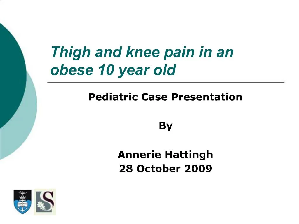
1. Thigh and knee pain in an obese 10 year old Pediatric Case Presentation
By
Annerie Hattingh
28 October 2009
2. History 10 yr old boy presents to ED with 1 week history of � thigh + knee pain.
He states that the pain is mainly in the thigh, but radiates down to his knee.
He was playing soccer when he collided with another player + fell.
Noted severe pain in his thigh + had to limp home on his left leg.
Since then, he has been complaining of pain in his � thigh when bearing weight.
3. History The pain would subside when lying down.
He did not improve much + was brought to ED.
He had no history of fever, rash, chest discomfort or pains in other joints.
4. Examination Vitals: Temp 37�C (oral)
PR 66
RR 20
BP 112/65
Weight: 59.3kg (>>95th percentile)
Height: 152cm (> 95th percentile)
Alert, cooperative + in no distress when lying down.
Obese + large for age.
5. Examination CVS: HR regular
(-) murmurs
Lungs: clear
Abdo: Round contour
Soft
Non-tender
6. Examination Musculoskeletal:
� lower extremity:
- Moderate tenderness in the upper ant.
thigh
- Severely tender hip, restricted ROM
- Pubic symphysis non tender
- Knee, tib-fib + foot non tender, normal
ROM
- No joint swelling noted
7. Examination Musculoskeletal:
(L) lower extremity:
- Mild tenderness of the hip on palpation
- Mild tenderness on ROM testing
- Rest unremarkable
NOTE: Although his chief complaint is thigh pain, the hip and knee joints should also be examined.
Hip injuries often present with knee pain.
8. Special investigations X-rays of the hips are ordered.
9. Diagnosis History of collision + fall suggests an acute injury such as a non-displaced #.
An obese child with hip pain in this age group should always raise the possibility of a SLIPPED FEMORAL EPIPHYSIS.
The X-ray shows a slipped capital femoral epiphysis on the �.
The left hip appears to be normal, however an early slip on the left is difficult to rule out.
10. Management He is hospitalized and bed rest ordered.
After a few hours of bed rest, his left hip is no longer tender.
He is referred to the orthopedic surgeon + taken to the OR for internal fixation of his � femoral epiphysis.
11. Discussion Radiographic dx of slipped femoral epiphysis can be subtle.
Clinical suspicion very important.
12. Discussion In this case, the physis appears to be wider + more lucent in the � hip, compared to the left.
13. Discussion The position of the femoral head epiphysis should resemble a cap over the physis.
Subtle cases may just show a slight malpositioning of the epiphysis.
14. Discussion Examine the following diagram of the pt�s hips:
15. Discussion The lines drawn along the superior border of the prox. femur metaphysis (the Klein line) should intersect part of the prox. femoral epiphysis.
16. Discussion The pt�s � hip ( left on the screen ) shows the line just touching the lateral margin of the epiphysis.
17. Discussion This is abnormal, indicating that the femoral epiphysis has slipped inferiorly + medially.
18. Discussion The normal left hip (right on the screen) shows the line intersecting the lateral part of the femoral epiphysis.
19. Discussion View this obvious case:
20. Discussion No line needs to be drawn here to appreciate that the pt�s left hip is abnormal.
Severe left slipped femoral epiphysis.
21. Discussion The slipped epiphysis on the � may not be so obvious.
Bilat. SFE is present, severe on the L + moderately severe on the R.
22. Discussion SCFE is a Dx that will occasionally present to the ED with an acute, sub acute or chronic pain in the hip, thigh or knee.
The Dx is not difficult if it is considered!!
Vague symptoms may be present
Degree of pain may range from severe to non-existent.
Ambulatory ability may range from non-weight bearing to normal gait.
23. Discussion Often wrongly diagnosed as:
- pulled muscle
- hip bruise
- hip/knee sprain
Patients tend to keep their hip externally rotated with inability to fully internally rotate the hip.
24. Discussion Risk factors:
Cause is unknown.
3-4 x more common in males than females.
Average age 10 � 16 years.
Pt�s are overweight for height/obese.
Associated with endocrine disorders like hypothyroidism, pituitary tumors + low growth hormone levels.
May be associated with minor fall or trauma
25. Discussion Radiographic diagnosis:
Obvious cases - epiphysis obviously displaced
Subtle cases - epiphyseal plate (physis) may be
widened / irregular compared to the other
side.
A line drawn along the sup. border of the metaphysis
( the Klein line ) may intersect less of the epiphysis compared to the normal side.
The epiphysis may appear to be thinner + occur if the slip is posteriorly.
26. Discussion Radiographic diagnosis:
Early slips difficult to detect on XR.
AP views only detect inferior + medial slips.
Posterior slips seen on lateral views ( but difficult to obtain ).
CT scans helpful to orthopedic surgeon - rarely done in Emergency Department.
MRI scanning not useful.
27. Discussion Treatment:
Responsibility of Orthopedic surgeon.
Prevent further slipping with internal fixation.
Important to make diagnosis on initial presentation!
Missed diagnosis may worsen slip and the future outcome.
28. Discussion Complications:
Avascular necrosis - most NB!
Premature osteoarthritis
Chondrolysis - loss of articular cartilage of the hip joint
- causes hip to stifffen with permanent loss
of motion, flexion contracture + pain.
29. The End
30. References MEDSCAPE pediatric trauma case studies
Online CME: Pediatrics
Rosen�s Emergency Medicine Online: Pediatric Trauma