Download
1 / 27

E N D
The strange glow emanating from Wilhelm Roentgen's laboratory in 1895 started out merely as part of an experiment to explore the laws of physics. But the invisible stream of particles producing this luminescence turned out to do a lot more. Roentgen found that the rays (which he labeled "x," since their properties were unknown) left impressions on photographic plates, making the seemingly solid and impenetrable–human skin and tissue–translucent and transparent. Within weeks, Roentgen's discovery had forever changed the understanding of human biology. "No one has ever seen the same way as they did before 1895," says Bettyann Holtzmann Kevles, a history lecturer at Yale University. As described in her book Naked to the Bone: Medical Imaging in the Twentieth Century, it was as if "the mind was walking in among the tissues themselves." In an era of bodily embarrassment, X-rays invited ordinary citizens to boldly reveal their inner selves. The cultural impact was stunning. Soon, X-ray machines for public entertainment appeared everywhere, although scientists later deemed them dangerous. In Chicago, people lined up to get a glimpse of the bones in their own hands at a coin-operated X-ray machine. At an opera house in Lawrence, Kan., couples could have X-ray snapshots taken as love tokens. And in New York, the wealthy visited X-ray studios to get their inner portraits done. Writers and artists embraced the unfettered spirit of the X-ray. In his 1897 novel, The Invisible Man, H. G. Wells built upon the already startling notion of seeing beneath the skin. His protagonist discovered rays that were similar to "Roentgen rays" but instead made the entire body invisible. A few years later, a group of painters in Italy who called themselves futurists adopted X-ray imagery in their early-20th-century manifesto. Asked their spokesman Umberto Boccioni, "Who can still believe in the opacity of bodies . . . ?" By then, X-rays had also become a medical necessity, proving critical for surgeons trying to set fractures and identify bullets or other foreign material in the body. "The X-ray served as a kind of beginning of a whole new way of delivering healthcare," says Joel Howell, professor of history and in?ternal medicine at the University of Michigan. In a departure from the patient's reliance on the family physician, "it helped build a profound and abiding trust in the idea that science and technology will lead to better healthcare," Howell adds. By the end of the 20th century, thanks to X-ray's daughter technologies–CAT, MRI, and PET– biological images took on sharp clarity, revealing not only bones but also muscle, tendons, blood vessels, and cartilage. Neurologists as well as scholars in philosophy, literature, and anthropology have taken to studying snapshots of the brain: What portions of the brain are affected by stroke? Where does our concept of self come from? How does love arise from our neural circuitry? And do novels activate the same synapses as daydreaming does? As scientists continue to pry deeply into the human body, they may one day illuminate–in the spirit of Roentgen–many more secrets of human nature.
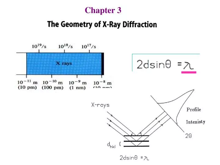
3.1 Generation of X-rays 1. Synchrotron X-rays
2. Tube X-rays X-rays For powder experiments X-rays For single-crystal experiments line focus spot focus in the plane perpendicular to the length of the filiment In the plane containing the length of the filiment
2. Tube X-rays X-rays X-rays If the electron loses all the energy, i.e. dropping through a potential V, then E = eV, and the minimum wavelength of the X-rays emitted will be = hc/E = 12398/V min (Å) ~12.4/KV White radiation is produced by simple collisions bw. electrons and target metal. If the electron loses all the energy, i.e. dropping through a potential V, then E = eV, and the minimum wavelength of the X-rays emitted will be = hc/E = 12398/V
wavelength Absorption spectrum at K edge The metal atoms in target may absorb the kinetic energy of the coming thermal electrons and get excited.
Characteristic X rays are dependent on the atomic character of the target material, generally used are copper and molybdenum. min ‘min Emission/characteristic spectrum of the target element min = 12398/V
Take-off angle 4o for fine-focus tube, 6o for normal-focus tube single-crystal XRD Powder XRD
3. X-rays from Rotating-Anode Generator The cathode rays are focused onto a line on the edge of the anode and the X-rays are taken off parallel to the plane of the disk. This system results in the generation of heat taking place over a considerable area-many times greater than the effective source area. disk with a bevelled edge (6-20o)
Using Metal Filter The absorption edge of Z-1 or Z-2 elements Target E
Kα aIn the above table, the thickness gives 50% reduction of Kαradiation. 1 Mil = 0.001 inch. The intensity of α1 line is twice that of α2, the average wavelength for the Kαlines can be calculated as λ(Kα) = [2λ(α1)+ λ(α2)]/3
Using monochromator monochromator
Exercise: Suppose that a graphite crystal (d002 = 3.348 Å) is used as a monochromator, (a) What will be the incident angles to get Cu K and Cu Kα1 lines? What will be it for Cu Kα2 line? (b) Do the same calculations as in (a) for Mo K, Mo Kα1 and Mo Kα2 lines. (c) What conclusions can be drawn from the results of (a) and (b)? The intensity of α1 line is twice that of α2, the average wavelength for the Kαlines can be calculated as λ(Kα) = [2λ(α1)+ λ(α2)]/3 _ 2 (Kα) = [2 (α1)+ 2 (α2)]/3
λ(α1) λ(α2) 2 (Kα) = [2 (α1)+ 2 (α2)]/3 At low angles : Exercise: The following PXRD diagram was recorded with Cu Kα1 radiation. If it were measured with the average Cu Kαlines, below what 2 angle will the Kα2 line will not be observed? (Assuming the resolution of the peaks is ~0.05o in 2 ).
Other Sources for diffraction measurements: Typical scattering curves for (1) electrons; (2) X-rays; and (3) neutrons Neutron diffraction analysis serves to locate hydrogen atoms (preferably as 2H) much more accurately than is possible by X-ray diffraction analysis. Neutron beam is often used in powder diffraction
Crystal Planes A plane intersecting the axes a, b, and c at the points m00,0n0,00p. The coordinates of the three intercepts completely define the position ofa lattice plane which is given a set of integer hkl, where a-axis: h = a/m; b-axis: k = b/n; c-axis: l = c/p
d100 = a d200 =½a dn00 =(1/n)a dnn0 = (1/n)d110 (100) (200) (110) Crystal Planes
5.628 Å Exercise: In the NaCl structure, both the Cl– (black dots) and the Na+ ions (open circles) form fcc. (i) Should the cell edge be 5.628 or 2.814 Å? (ii) How many lattice points are in an unit cell? (iii) Assign the crystal axes and define the three sets of lattice planes in the following figures.
diffraction space a reflection (diffraction peak) crystal space
General equations used for d-spacing • 2dhk sinθhk =λ • Qhk = d-2hk = (4/λ2) sin2θhk • dhk- 2 = ha*2 + k2b*2 + c*2 +2kb*c*cos* • + 2 hc*a*cos* + 2 hka*b*cos* • d-2hk =h2A + k2B + 2C + hkD + hE + kF • (a) Cubic: d-2 = (h2+k2+ 2)A (1 parameter) • (b) Hexagonal: d-2 = (h2+hk+k2)A + 2C (2 parameters) • (c) Tetragonal: d-2 = (h2+k2)A + 2C • (d) Orthorhombic: d-2 = h2A+k2B+ 2C (3 parameters) • (e) Monoclinic: d-2 = h2A+k2B+ 2C+hE (4 parameters)
Hexagonal Axes vs. Rhombohedral Axes Two ways to relate rhombohedral indices to hexagonal indices, the obverse and reverse relationship. The hexagonal cell : a1, a2, c and indices (h k .) Rhombohedral cell: r1, r2 , r3and indices (m n p) Obversehexagonal axes: a1 = r2 – r3 a2 = r3 – r1 c = r1 + r2+ r3 h = n – p k = -m + p = m + n + p -h + k + = 3p Reverse hexagonal axes: a1 = r3 – r2 a2 = r1 – r3 c = r1 + r2+ r3 h - k + = 3p Obverse H vs. R: